. 2018 May 1;159(5):2142-2152.
doi: 10.1210/en.2018-00017.

Multiple cAMP Phosphodiesterases Act Together to Prevent Premature Oocyte Meiosis and Ovulation

Affiliations

Multiple cAMP Phosphodiesterases Act Together to Prevent Premature Oocyte Meiosis and Ovulation

Giulia Vigone et al. Endocrinology. .

Abstract

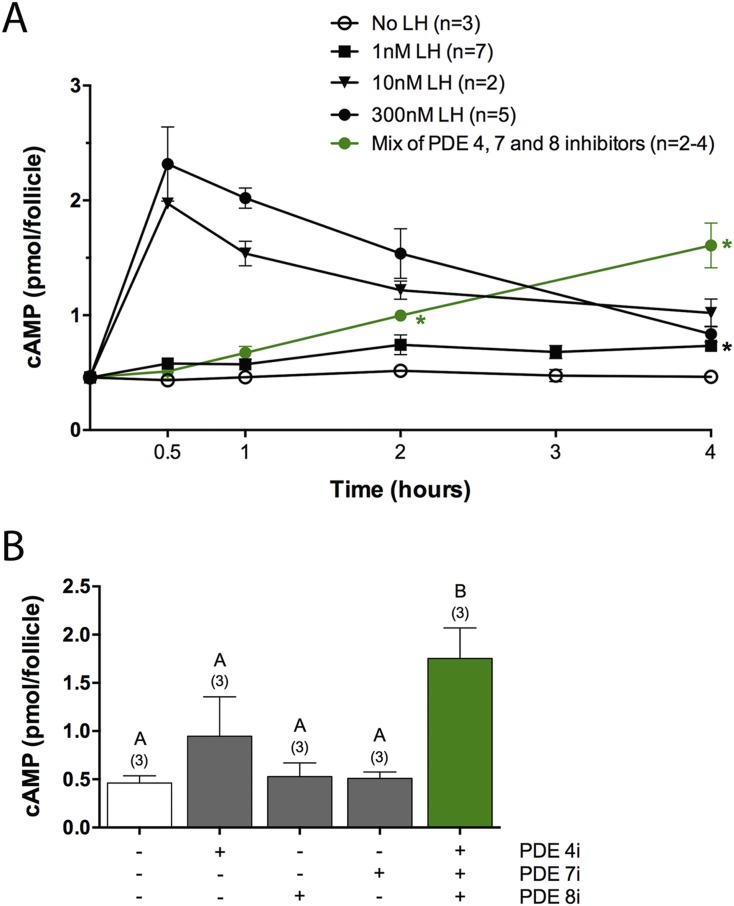
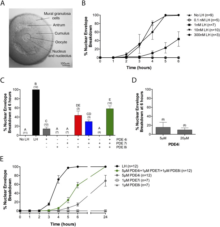
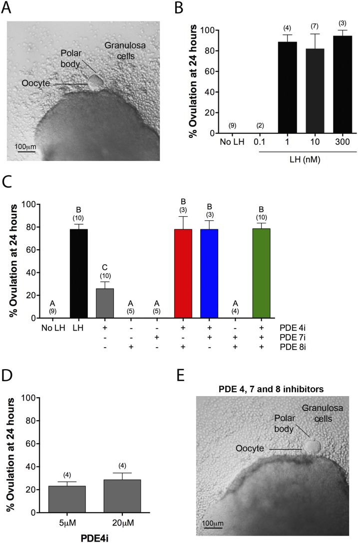
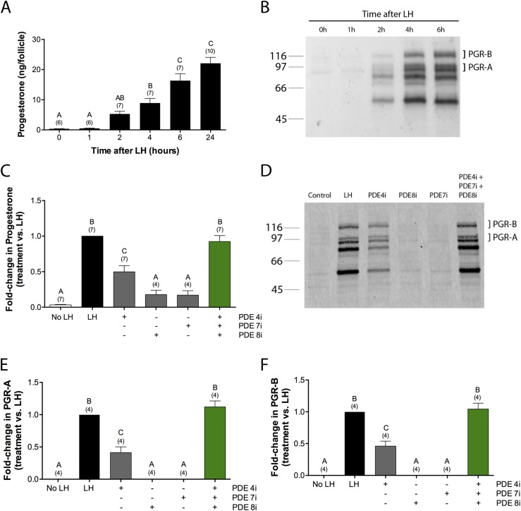
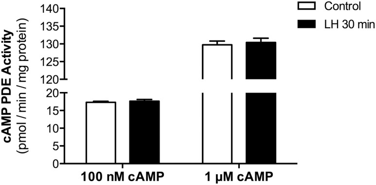
Luteinizing hormone (LH) acts on the granulosa cells that surround the oocyte in mammalian preovulatory follicles to cause meiotic resumption and ovulation. Both of these responses are mediated primarily by an increase in cyclic adenosine monophosphate (cAMP) in the granulosa cells, and the activity of cAMP phosphodiesterases (PDEs), including PDE4, contributes to preventing premature responses. However, two other cAMP-specific PDEs, PDE7 and PDE8, are also expressed at high levels in the granulosa cells, raising the question of whether these PDEs also contribute to preventing uncontrolled activation of meiotic resumption and ovulation. With the use of selective inhibitors, we show that inhibition of PDE7 or PDE8 alone has no effect on the cAMP content of follicles, and inhibition of PDE4 alone has only a small and variable effect. In contrast, a mixture of the three inhibitors elevates cAMP to a level comparable with that seen with LH. Correspondingly, inhibition of PDE7 or PDE8 alone has no effect on meiotic resumption or ovulation, and inhibition of PDE4 alone has only a partial and slow effect. However, the fraction of oocytes resuming meiosis and undergoing ovulation is increased when PDE4, PDE7, and PDE8 are simultaneously inhibited. PDE4, PDE7, and PDE8 also function together to suppress the premature synthesis of progesterone and progesterone receptors, which are required for ovulation. Our results indicate that three cAMP PDEs act in concert to suppress premature responses in preovulatory follicles.


Conflict of interest statement

The authors have nothing to disclose.

Figures

Figure 1.
Figure 1.
PDE4, PDE7, and PDE8 act together to suppress premature cAMP elevation in preovulatory follicles. (A) Time course of cAMP elevation after exposure of follicles to various concentrations of LH or to a mixture of inhibitors of three cAMP PDEs: PDE4i (rolipram, 5 μM), PDE7i (1 μM), and PDE8i (PF04957325, 1 μM). The LH data were obtained from the indicated number of independent experiments, each including all time points. The PDE inhibitor data include two to four measurements for each time point. t Tests were performed to test the statistical significance of the difference between the measurements following no LH and 1 nM LH treatments; the black asterisk indicates a significant difference (P < 0.05 after correction for multiple comparisons). t Tests were also used to test the difference between measurements following no LH and PDE inhibitor treatments; the green asterisks indicate significant differences. (B) cAMP content of follicles after a 4-hour exposure to each of the inhibitors listed in (A) or to a mixture of the three inhibitors. Numbers in parentheses indicate the number of independent experiments. Different letters indicate significant differences (P < 0.05).
Figure 2.
Figure 2.
PDE4, PDE7, and PDE8 act together to suppress premature NEBD in follicle-enclosed oocytes. (A) A follicle before exposure to LH or a PDE inhibitor; an intact nucleus is visible in the oocyte. (B) Time course of NEBD in response to various concentrations of LH. (C) Percent NEBD at 6 hours after treatment of follicles with a saturating concentration of LH (300 nM), individual PDE inhibitors, pairs of inhibitors, or all three inhibitors together (5 μM rolipram, 1 μM PDE7i, 1 μM PDE8i). (D) Similar percent NEBD in response to 5 and 20 μM rolipram. (E) Time course of NEBD in response to LH (300 nM), individual PDE inhibitors [concentrations as in (C)], or a mixture of the three inhibitors. Numbers in parentheses indicate the number of independent experiments. Different letters indicate significant differences (P < 0.05).
Figure 3.
Figure 3.
PDE4, PDE7, and PDE8 act together to suppress premature ovulation. (A) A follicle at 24 hours after exposure to LH (300 nM). Ovulation has occurred, as indicated, by extrusion of the oocyte and some granulosa cells. (B) Percent ovulation at 24 hours after exposure to various concentrations of LH. (C) Percent ovulation at 24 hours after treatment of follicles with LH (300 nM), individual PDE inhibitors, pairs of inhibitors, or all three inhibitors together (5 μM rolipram, 1 μM PDE7i, 1 μM PDE8i). (D) Similar percent ovulation in response to 5 and 20 μM rolipram. (E) A follicle at 24 hours after treatment with a mixture of PDE4i, PDE7i, and PDE8i [concentrations as in (C)], showing ovulation, as seen with LH. Numbers in parentheses indicate the number of independent experiments. Different letters indicate significant differences (P < 0.05).
Figure 4.
Figure 4.
PDE4, PDE7, and PDE8 all contribute to suppression of spontaneous production of progesterone and PGRs. (A and B) Time course of synthesis of (A) progesterone and (B) its receptors, PGR-A and PGR-B, in response to LH (300 nM). (C–F) Stimulation of synthesis of (C) progesterone and (D–F) PGRs by incubation of follicles for 6 hours in the presence of LH (300 nM) or PDE4i (rolipram, 5 μM), PDE7i (1 μM), and/or PDE8i (1 μM). (B and D) Molecular weight in kDa is shown at left. The doublet near the 97 kDa marker corresponds to the PGR-A isoform, and the doublet just above the 116 kDa marker corresponds to the PGR-B isoform; the doublets are thought to result from phosphorylation (32). The lower bands, in the range of ∼50 to 70 kDa, are unidentified but have been seen in a previous study as well (32). Numbers in parentheses indicate the number of independent experiments. Different letters indicate significant differences (P < 0.05).
Figure 5.
Figure 5.
LH signaling does not cause a rapid decrease in cAMP PDE activity. cAMP PDE activity was measured in lysates of follicles, with or without a 30-minute exposure to LH (300 nM). Activity was assayed with two different cAMP substrate concentrations: 0.1 or 1 μM. Bars show the means ± SEM for two independent experiments.

Similar articles

Cited by

References

    1. Conti M, Kasson BG, Hsueh AJ. Hormonal regulation of 3′,5′-adenosine monophosphate phosphodiesterases in cultured rat granulosa cells. Endocrinology. 1984;114(6):2361–2368. - PubMed
    1. Tsafriri A, Chun SY, Zhang R, Hsueh AJ, Conti M. Oocyte maturation involves compartmentalization and opposing changes of cAMP levels in follicular somatic and germ cells: studies using selective phosphodiesterase inhibitors. Dev Biol. 1996;178(2):393–402. - PubMed
    1. Lyga S, Volpe S, Werthmann RC, Götz K, Sungkaworn T, Lohse MJ, Calebiro D. Persistent cAMP signaling by internalized LH receptors in ovarian follicles. Endocrinology. 2016;157(4):1613–1621. - PubMed
    1. Zhang M, Su YQ, Sugiura K, Xia G, Eppig JJ. Granulosa cell ligand NPPC and its receptor NPR2 maintain meiotic arrest in mouse oocytes. Science. 2010;330(6002):366–369. - PMC - PubMed
    1. Hunzicker-Dunn M, Mayo K. Gonadotrophin signaling in the ovary In: Plant TM and Zeleznik AJ, eds. Knobil and Neill’s Physiology of Reproduction. 4th ed.San Diego: Academic Press; 2015:895–945.

Publication types

MeSH terms

Substances

LinkOut - more resources