. 2018 Aug;97(9):987-994.
doi: 10.1177/0022034518765757. Epub 2018 Apr 2.

Biomechanics of Immediate Postextraction Implant Osseointegration

Affiliations

Biomechanics of Immediate Postextraction Implant Osseointegration

X Yuan et al. J Dent Res. 2018 Aug.

Abstract

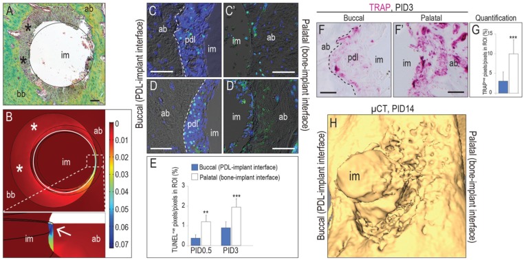
The aim of this study was to gain insights into the biology and mechanics of immediate postextraction implant osseointegration. To mimic clinical practice, murine first molar extraction was followed by osteotomy site preparation, specifically in the palatal root socket. The osteotomy was positioned such that it removed periodontal ligament (PDL) only on the palatal aspect of the socket, leaving the buccal aspect undisturbed. This strategy created 2 distinct peri-implant environments: on the palatal aspect, the implant was in direct contact with bone, while on the buccal aspect, a PDL-filled gap existed between the implant and bone. Finite element modeling showed high strains on the palatal aspect, where bone was compressed by the implant. Osteocyte death and bone resorption predominated on the palatal aspect, leading to the loss of peri-implant bone. On the buccal aspect, where finite element modeling revealed low strains, there was minimal osteocyte death and robust peri-implant bone formation. Initially, the buccal aspect was filled with PDL remnants, which we found directly provided Wnt-responsive cells that were responsible for new bone formation and osseointegration. On the palatal aspect, which was devoid of PDL and Wnt-responsive cells, adding exogenous liposomal WNT3A created an osteogenic environment for rapid peri-implant bone formation. Thus, we conclude that low strain and high Wnt signaling favor osseointegration of immediate postextraction implants. The PDL harbors Wnt-responsive cells that are inherently osteogenic, and if the PDL tissue is healthy, it is reasonable to preserve this tissue during immediate implant placement.

Keywords: Wnt3 protein; cell lineage; dental implantation; osteogenic; periodontal ligament; tooth extraction.


Conflict of interest statement

The authors declare no potential conflicts of interest with respect to the authorship and/or publication of this article.

Figures

Figure 1.
Figure 1.
Around immediate postextraction implants, gap interfaces constitute a low-strain pro-osteogenic environment. (A) Per histologic examination on postimplant day 0.5 (PID0.5), at the palatal site, the immediate implant (black circle) surface was in direct contact with bone, and at the buccal site, the gap between alveolar bone and implant contained residual PDL fibers (asterisks). (B) Magnitude of the strain in the peri-implant bone according to finite element modeling. White arrow points to the highest strain area. On PID0.5, (C) on buccal aspects of the implants (PDL-implant interface), TUNEL+ve cells were restricted in the PDL remnants, (C′) whereas on the palatal aspects (bone-implant interface), TUNEL staining identified many osteocytes undergoing programmed cell death as a result of osteotomy site preparation and implant placement (quantified in E). On PID3, (D) on the buccal aspects of the implants, TUNEL+ve cells was found only in the PDL remnants, whereas (D′) TUNEL+ve osteocytes were extended ~100 µm from the osteotomy edge on the palatal aspects (quantified in E). As a consequence, (F) minimal TRAP activity was found on the buccal sites, which was restricted to the PDL remnants. (F′) On palatal aspects, TRAP activity was detectable throughout in the bone (quantified in G). (H) On PID14, micro–computed tomography (μCT) showed significant crestal recession on the palatal surfaces where the implant was in direct contact with bone. The dashed line (C, D, F) indicates the demarcation between alveolar bone and PDL remnants. (E, G) Values are presented as mean ± SD. ab, alveolar bone; bb, bundle bone; im, implant; pdl/PDL, periodontal ligament; ROI, region of interest. Scale bars = 50 µm. **P < 0.01. ***P < 0.0001.
Figure 2.
Figure 2.
Osteoprogenitor cells residing in the periodontal ligament (PDL) support peri-implant bone formation. (A) Schematic of implant in the palatal root. On the buccal aspect of the implant, a gap filled with PDL exists between the alveolar bone and the implant (PDL-implant interface), and on the palatal aspect of the implant, the alveolar bone is directly contact with the implant (bone-implant interface). On postimplant day 7 (PID7), (B) buccal aspects with residual PDL showed new bone formed at the interface, whereas (B′) palatal aspects only displayed fibrous interfacial tissue. (C) Buccal aspects consistently showed robust ALP activity as compared with (C′) palatal aspects. (D) Buccal aspects of implants largely comprised osterix+ve osteoblasts, whereas (D′) palatal aspects had fewer osterix+ve cells. Type I collagen staining indicates bone matrix deposited (E) on the buccal aspects but not (E′) the palatal aspects. By PID14, (F) buccal surfaces of implants were surrounded by mature bone, whereas (F′) on palatal aspects of implants, new bone formation just started. Picrosirius red staining shows mature bone formed (G) on the buccal aspects but not (G′) the palatal aspects. (H) The buccal aspects of implants largely comprised mature bone with osteocalcin+ve osteoblasts and osteocytes, while (H′) fewer osteocalcin+ve cells were found on the palatal aspects. Calcein (green, labeling the existed alveolar bone) and alizarin red (red, labeling the newly formed bone) assay showed that (I) peri-implant bone formed on the buccal aspects, whereas (I′) new bone formed around the existed alveolar bone on the palatal aspects. The red dotted line indicates the edge of existed alveolar bone; the white dashed line indicates the edge of implant. Scale bars = 50 µm for all panels. ALP, alkaline phosphatase; OCN, osteocalcin.
Figure 3.
Figure 3.
Progeny of a Wnt-responsive population is responsible for peri-implant bone formation. (A) GFP+ve Wnt-responsive cells in the mxM1 PDL of 4-wk-old mice 5 d after tamoxifen treatment. (B) On postimplant day 3 (PID3), similar GFP+ve cell patterns were found around immediate the postextraction implants. In the intact PDL, those Wnt-responsive PDL cells were (C) periostin+ve, but most of them were (E) absent of Ki67 expression. On PID3, GFP+ve cells were (D) periostin+ve but (F) highly proliferative as evidenced by Ki67 expression. On PID7, the buccal aspects were dominated by (G) GFP+ve cells expressing (H) osterix and (I) osteocalcin. At the same time point, on the palatal aspects of the same implants, only a few (G′) GFP+ve, (H′) osterix+ve, and (I′) osteocalcin+ve cells were detected. By PID14, the buccal aspects of the implant comprised mature bone with (J) GFP+ve cells that were (K) osteocalcin+ve and aligning on the newly formed bone surface or trapped inside. On the palatal aspects of the implant, (J′) GFP+ve cells and (K′) osteocalcin+ve cells were in evidence but not surrounded by bone matrix yet. Pentachrome staining showing the mature bone on (L) the buccal aspects but not (L′) the palatal aspects. Abbreviations as noted in Figure 1. d, dentin. The dotted line indicates edge of PDL; the dashed line indicates the edge of implant. Scale bars = 50 µm for all panels.
Figure 4.
Figure 4.
Wnt signaling accelerates peri-implant bone formation. On the palatal aspects of implants, (A) the liposomal WNT3A (L-WNT3A) treatment group showed more Xgal+ve Wnt-responsive cells as compared with the (A′) liposomal phosphate-buffered saline (L-PBS; control) group on postimplant day 3 (PID3). The L-WNT3A group showed (B, B′) minimal osteocyte apoptosis as well as (C, C′) TRAP activity as compared with the L-PBS group. (D, D′) The L-WNT3A-treated group also displayed stronger ALP activity. On PID7, L-WNT3A treatment promoted (E, E′) type I collagen and (F, F′) RUNX2 expression, therefore, (G, G′) showed peri-implant bone formation. (H) The evaluation of bone-implant contact (BIC). Values are presented as mean ± SD. ***P < 0.0001. On the buccal aspects of implants, the L-WNT3A group showed more (I, I′) Xgal+ve Wnt-responsive cells and (J, J′) RUNX2+ve cells on PID3. (K, K′) On PID7, the L-WNT3A group showed mature peri-implant bone formation. (L, L′) Micro–computed tomography showed superior peri-implant bone formation in the L-WNT3A group on both buccal and palatal aspects. Abbreviations as noted in Figure 1. ALP, alkaline phosphatase; BIC, bone-implant contact. Scale bars = 50 µm for all panels. The dashed line indicates the tissue-implant interface.

References

    1. Almeida M, Han L, Bellido T, Manolagas SC, Kousteni S. 2005. Wnt proteins prevent apoptosis of both uncommitted osteoblast progenitors and differentiated osteoblasts by beta-catenin-dependent and -independent signaling cascades involving Src/ERK and phosphatidylinositol 3-kinase/AKT. J Biol Chem. 280(50):41342–41351. - PubMed
    1. Anitua E, Pinas L, Alkhraisat MH. 2016. Long-term outcomes of immediate implant placement into infected sockets in association with immediate loading: a retrospective cohort study. J Periodontol. 87(10):1135–1140. - PubMed
    1. Bonewald LF, Johnson ML. 2008. Osteocytes, mechanosensing and Wnt signaling. Bone. 42(4):606–615. - PMC - PubMed
    1. Branemark PI, Hansson BO, Adell R, Breine U, Lindstrom J, Hallen O, Ohman A. 1977. Osseointegrated implants in the treatment of the edentulous jaw: experience from a 10-year period. Scand J Plast Reconstr Surg Suppl. 16:1–132. - PubMed
    1. Brunski JB. 1999. In vivo bone response to biomechanical loading at the bone/dental-implant interface. Adv Dent Res. 13:99–119. - PubMed

Publication types

LinkOut - more resources