Review
. 2018 Nov 1:127:248-261.
doi: 10.1016/j.freeradbiomed.2018.03.051. Epub 2018 Mar 30.

NADPH-dependent and -independent disulfide reductase systems

Affiliations
Review

NADPH-dependent and -independent disulfide reductase systems

Colin G Miller et al. Free Radic Biol Med. .

Abstract

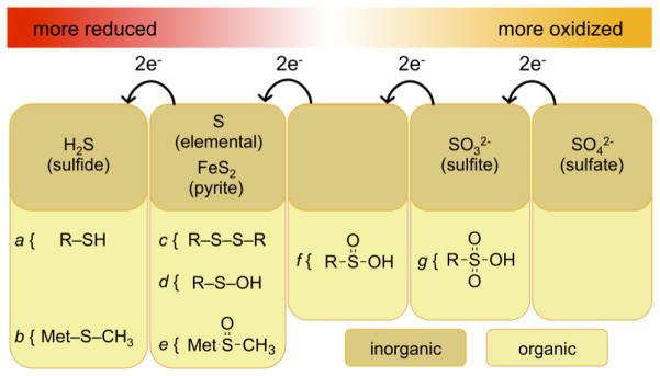
Over the past seven decades, research on autotrophic and heterotrophic model organisms has defined how the flow of electrons ("reducing power") from high-energy inorganic sources, through biological systems, to low-energy inorganic products like water, powers all of Life's processes. Universally, an initial major biological recipient of these electrons is nicotinamide adenine dinucleotide-phosphate, which thereby transits from an oxidized state (NADP+) to a reduced state (NADPH). A portion of this reducing power is then distributed via the cellular NADPH-dependent disulfide reductase systems as sequential reductions of disulfide bonds. Along the disulfide reduction pathways, some enzymes have active sites that use the selenium-containing amino acid, selenocysteine, in place of the common but less reactive sulfur-containing cysteine. In particular, the mammalian/metazoan thioredoxin systems are usually selenium-dependent as, across metazoan phyla, most thioredoxin reductases are selenoproteins. Among the roles of the NADPH-dependent disulfide reductase systems, the most universal is that they provide the reducing power for the production of DNA precursors by ribonucleotide reductase (RNR). Some studies, however, have uncovered examples of NADPH-independent disulfide reductase systems that can also support RNR. These systems are summarized here and their implications are discussed.

Keywords: Cysteine; Glutathione reductase; Methionine cycle; NADPH; Ribonucleotide reductase; Sulfur amino acid; Thioredoxin reductase; Transsulfuration.


Figures

Fig. 1
Fig. 1. Chemical structure and activity of the [NADPH;NADP+] redox couple
The redox-active electrons are denoted in red font by the symbol e. The boxed inset shows the structural difference between the NADP- and the NAD-family of nicotinamide cofactors.
Fig. 2
Fig. 2. Oxidation states of common inorganic and organic S-compounds
For simplicity, only species discussed in this review are included. Organic compounds are as follows: a, thiols, such as found in reduced proteins, Cys, and GSH; b, thioether within Met; c, disulfide, such as found in oxidized protein-disulfides or GSSG; d, sulfenic acid; e, sulfoxide within oxidized Met; f, sulfinic acid; g, sulfonic acid.
Fig. 3
Fig. 3. Alternative pathways of E. coli disulfide reduction
A, constitutive disulfide reduction by NADPH-dependent TrxR- or Gsr-driven systems. B, in TrxR/Gsr-null E. coli, a suppressor mutation in the AhpC gene (AhpC*) results in the enzyme being able to use reducing power from AhpF to reduce Grx1-SSG. AhpF can use either NADPH or NADH as electron donors, so this provides a possible source of NADHP-independent disulfide reducing power. C, left, normal TCA cycle drives reduction of LA-disulfide cofactors to the dihydrolipoate state and LpdA uses this reducing power to drive reduction of NAD+ → NADH. The NADH is used for bioenergetics and does not contribute to disulfide reduction. Right, in triple-null TrxR/Gsr/AhpC mutants, suppressor mutations (LpdA*) compromise LpdA activity. Accumulated reducing power on the dihydrolipoate cofactors is then transferred to oxidized Grx-disulfide, generating Grx-dithiol. This regenerates the LA-disulfide, thereby restoring TCA cycle activity, and allows Grx to provide reducing power to RNR. This system is fully NADPH-independent.
Fig. 4
Fig. 4. Met metabolism and transsulfuration pathway activities
A, acquisition of disulfide reducing power from extracellular Met. The redox-active S is designated in red font as S. In extracellular fluids, the reduced S is protected from oxidation as a thioether. The Met-cycle generates Hcy bearing this reduced S, and transsulfuration moves this reduced S to Cys; no other portion of Met is transferred to the Cys. The Cys can be used in de novo GSH biosynthesis, which puts the reduced S into a context that can be used for disulfide reduction reactions. The points of pathway inhibition by PPG and BSO are shown. Ancillary reactants and products are not shown. B, Roles of CBS and CSE in S-amino acid homeostasis and in H2S signaling. Row 1 shows the classical transsulfuraion reactions for shunting S from the Met-cycle into Cys. Row 2 shows the reactions that lead to H2S production for signaling. For the CBS reaction, Cys replaces Ser, so the reaction is Hcy + Cys → cystathionine + H2S. For the Cse reaction, cystine replaces cystathionine, so the reaction is cystine → thiocystine + pyruvate. In a subsequent step not shown, thiocystine can be reduced by TRP14, Trx1, or Grx to generate H2S + Cys.
Fig. 5
Fig. 5. Cellular metabolism of sulfur
Inorganic S metabolism, de novo production of Cys using sulfide, and de novo synthesis of Met from Cys are all restricted to plants and microbes. Metazoans have reversed the transsulfuration pathway, and thereby use Met for de novo synthesis of Cys. Scheme shows only routes and directions, not redox requirements at each step.
Fig. 6
Fig. 6. Phylogeny of TrxR and Gsr enzyme families, and of Met auxotrophy
A.t. – Arabidopsis thaliana, A.q. – Amphimedon queenslandica, C.e. – Caenorhabitis elegans, C.i. – Ciona intestinalis, C.r. – Chlamydomonas reinhardtii, D.d. – Dictyostelium discoideum, D.m. – Drosophila melanogaster, E.c. – Escherichia coli, H.s. – Homo sapiens, M.m. – Mus musculus, S.c. – Saccharomyces cerevisiae. The asterisk denotes the single-celled green alga, C. reinhardtii, as representing a sparse but diverse set of non-metazoans that have the metazoan TrxR (blue line with “?”). Predominant others in this group are protozoan intracellular parasites, which might have acquired the gene laterally from a metazoan host. To our knowledge, no similar model yet explains the enigmatic presence of this gene in an alga.

References

    1. Buchanan BB. Regulation of CO2 assimilation in oxygenic photosynthesis: the ferredoxin/thioredoxin system. Perspective on its discovery, present status, and future development. Arch Biochem Biophys. 1991;288(1):1–9. - PubMed
    1. Calvin M, Benson AA. The Path of Carbon in Photosynthesis. Science. 1948;107(2784):476–80. - PubMed
    1. Bassham JA, Benson AA, Kay LD, Harris AZ, Wilson AT, Calvin M. The Path of Carbon in Photosynthesis. XXI. The Cyclic Regeneration of Carbon Dioxide Acceptor1. Journal of the American Chemical Society. 1954;76(7):1760–1770.
    1. Fan J, Ye J, Kamphorst JJ, Shlomi T, Thompson CB, Rabinowitz JD. Quantitative flux analysis reveals folate-dependent NADPH production. Nature. 2014;510(7504):298–302. - PMC - PubMed
    1. Margittai É, Bánhegyi G. Isocitrate dehydrogenase: A NADPH-generating enzyme in the lumen of the endoplasmic reticulum. Archives of Biochemistry and Biophysics. 2008;471(2):184–190. - PubMed

Publication types