. 2018 Jun:168:24-37.
doi: 10.1016/j.biomaterials.2018.03.044. Epub 2018 Mar 26.

Heparin-Poloxamer Thermosensitive Hydrogel Loaded with bFGF and NGF Enhances Peripheral Nerve Regeneration in Diabetic Rats

Affiliations

Heparin-Poloxamer Thermosensitive Hydrogel Loaded with bFGF and NGF Enhances Peripheral Nerve Regeneration in Diabetic Rats

Rui Li et al. Biomaterials. 2018 Jun.

Abstract

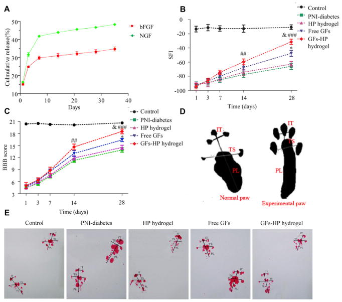
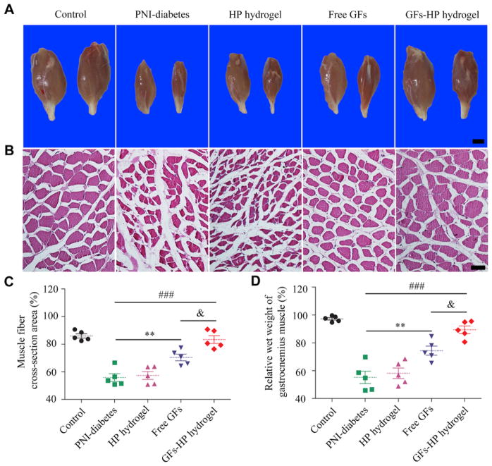
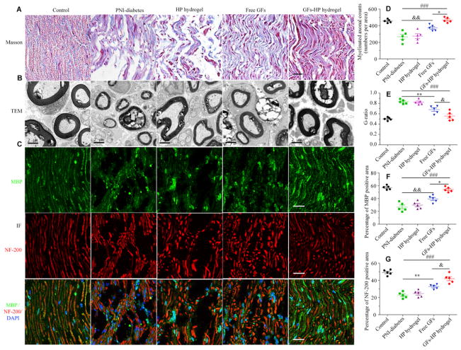
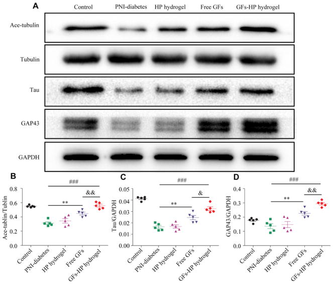
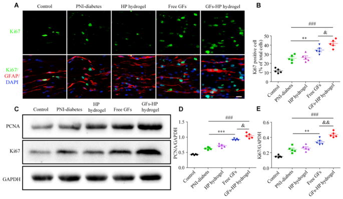
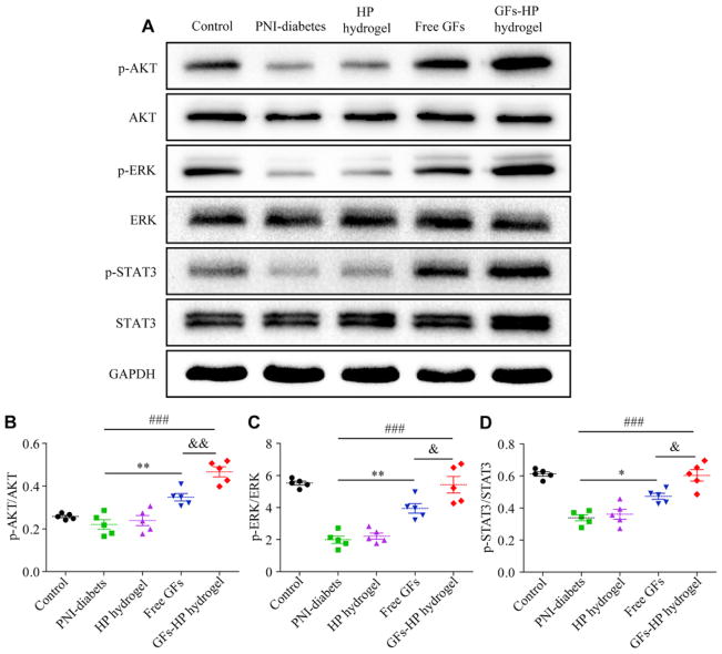
Peripheral nerve injury (PNI) is a major burden to society with limited therapeutic options, and novel biomaterials have great potential for shifting the current paradigm of treatment. With a rising prevalence of chronic illnesses such as diabetes mellitus (DM), treatment of PNI is further complicated, and only few studies have proposed therapies suitable for peripheral nerve regeneration in DM. To provide a supportive environment to restore structure and/or function of nerves in DM, we developed a novel thermo-sensitive heparin-poloxamer (HP) hydrogel co-delivered with basic fibroblast growth factor (bFGF) and nerve growth factor (NGF) in diabetic rats with sciatic nerve crush injury. The delivery vehicle not only had a good affinity for large amounts of growth factors (GFs), but also controlled their release in a steady fashion, preventing degradation in vitro. In vivo, compared with HP hydrogel alone or direct GFs administration, GFs-HP hydrogel treatment is more effective at facilitating Schwann cell (SC) proliferation, leading to an increased expression of nerve associated structural proteins, enhanced axonal regeneration and remyelination, and improved recovery of motor function (all p < 0.05). Our mechanistic investigation also revealed that these neuroprotective and neuroregenerative effects of the GFs-HP hydrogel may be associated with activations of phosphatidylinositol 3 kinase and protein kinase B (PI3K/Akt), janus kinase/signal transducer and activator of transcription 3 (JAK/STAT3), and mitogen-activated protein kinase kinase/extracellular signal-regulated kinase (MAPK/ERK) signaling pathways. Our work provides a promising therapy option for peripheral nerve regeneration in patients with DM.

Keywords: Basic fibroblast growth factor; Diabetes; Heparin-poloxamer; Nerve growth factor; Nerve regeneration; Peripheral nerve injury.


Conflict of interest statement

Conflicts of interest

The authors state no conflict of interest.

Figures

Fig. 1
Fig. 1
The thermosensitive property of GFs-HP hydrogel. A, C. Visualization of the state of HP and GFs-HP respectively at different temperatures (4°C, 37°C and 4°C after 37°C). At 37°C, both hydrogels are clearly in a gel state, while at 4°C before or after heating, both hydrogels are clearly in a sol state. B, D. Storage (G′) and loss (G″) moduli as rheological markers in temperatures ranging from 10 to 40°C for HP and GFs-HP hydrogels, respectively. Results show that the temperature of gel-sol phase transition for HP is slightly higher than that of GFs-HP, but both are well below 37°C.
Fig. 2
Fig. 2
The microstructure and safety of HP-hydrogel with/without GFs. A. SEM image of the morphology HP and GFs-HP hydrogel. Scale bar=100μm; B. The survival rate of RSC96cells treated with HP with/without GFs using PI/annexin V-FITC staining and statistical results of early apoptosis rate. Results show that there is no appreciable change in apoptotic rate of SCs with GFs-HP hydrogel. n.s, nonsignifcant; C. The stability of the bFGF and NGF in the hydrogel at different times was evaluated by CCK-8 assay. All data represent mean values±SEM, n=3 for each group.
Fig. 3
Fig. 3
The continuous release of bFGF and NGF from GFs-HP hydrogel enhances motor functional recovery of the regenerated sciatic nerve. A. Release profile of encapsulated bFGF and NGF at different time points; B. The sciatic function index (SFI) values in all groups measured at the predetermined time postoperatively; C. Quantification of BBB scores in the indicated groups at the predetermined time postoperatively; D. A schematic for measuring parameters of footprints; E. Representative photographs of the rats’ paw prints in each group 28 days after sciatic nerve crush injury. Free GFs vs PNI-diabetes: *P <0.05, GFs-HP hydrogel vs PNI-diabetes: ##P <0.01, ###P <0.001, GFs-HP hydrogel vs Free GFs: &P <0.05. All data represent mean values±SEM, n=8 in each group.
Fig. 4
Fig. 4
Analyses gastrocnemius muscle morphology and wet weight for evaluation of post-injury muscle atrophy. A, Representative photographs of gastrocnemius muscles from both hind limbs in each group. Scale bar, 25mm; B, Representative light microscopy images of cross-sectioned gastrocnemius muscles following H&E staining 30 days post-injury. Scale bar, 50μm; C. Histograms showing the percentage of cross-sectional area of muscle fibers quantified with Image-Pro Plus software analysis of light microscopy results; D. Wet weight ratios of gastrocnemius muscles in each group at 30 days post injury. Free GFs vs PNI-diabetes: **P <0.01, GFs-HP hydrogel vs PNI-diabetes: ###P <0.001, GFs-HP hydrogel vs Free GFs: &P <0.05. All data represent mean values±SEM, n=5 for each group.
Fig. 5
Fig. 5
Histological and microstructure evaluation of injured sciatic nerve. A. longitudinal sections of regenerated nerve stained with Masson’s trichrome 30 days after injury, Scale bar=50μm; B. TEM images of cross-sections of lesion regions, Scale bar=2μm; C. Double immunofluorescence staining for NF-200 and MBP-positive cells of the longitudinal sections in each experimental group. Scale bar=25μm; D, E. Quantification analysis of myelinated axonal count and G-ratio (G-ratio=axon diameter/fiber diameter) in the indicated groups; F, G. Quantitative analyses of fluorescence intensity of pixels for MBP and NF-200. Values are expressed as mean±SEM, n=5 per group. Free GFs vs PNI-diabetes: *P <0.05, **P <0.01, GFs-HP hydrogel vs PNI-diabetes: ###P <0.001, GFs-HP hydrogel vs Free GFs: &P <0.05, &&P <0.01.
Fig. 6
Fig. 6
GFs-HP hydrogel maintains MT stability in axons and upregulates GAP43 expression. A. the expression levels of MT-associated proteins (Ace-tubulin, Tyr-tubulin, Tau) and neural structural protein GAP43 in sciatic nerve lesions from diabetic rats via western blotting (GAPDH served as a protein loading control); B, C, D. Densitometrical analysis of Ace-tubulin, Tyr-tubulin, Tau and GAP43, respectively. Data presented as mean±SEM, n=5 for each group. Free GFs vs PNI-diabetes: **P <0.01, GFs-HP hydrogel vs PNI-diabetes: ###P <0.001, GFs-HP hydrogel vs Free GFs: &P <0.05, &&P <0.01.
Fig. 7
Fig. 7
GFs-HP hydrogel facilitates SCs proliferation. A. The double immunofluorescence staining of Ki67 (green) with the GFAP (red) to label proliferating SCs in longitudinal sciatic nerve sections, Scale bar=50μm; B. The percentages of cells double-positive for Ki67 and GFAP out of total DAPI positive cells (representing proliferation rate of SCs); C. The protein levels of Ki67 and PCNA at 30 days after injury by western blotting; D, E. Densitometric analyses of PCNA and Ki67, respectively. GAPDH was used for band density normalization. Data presented as mean±SEM, n=5 for each group. Free GFs vs PNI-diabetes: **P <0.01, ***P <0.001, GFs-HP hydrogel vs PNI-diabetes: ###P<0.001, GFs-HP hydrogel vs Free GFs: &P <0.05, &&P <0.01. (For interpretation of the references to color in this figure legend, the reader is referred to the Web version of this article.)
Fig. 8
Fig. 8
GFs-HP hydrogel treatment activates SCs proliferation through MAPK/ERK, PI3K/Akt and JAK/STAT3 pathways. A. Immunoblot for p-AKT, p-ERK, p-STAT3; total proteins amount of AKT, ERK, STAT3 served as loading control; B–D: Densitometric quantification of p-AKT/AKT, p-ERK/ERK and p-STAT3/STAT3, respectively. GAPDH was used for band density normalization. Data presented as Mean±SEM n=5 for each group. Significance markers: Free GFs vs PNI-diabetes: *P <0.05, **P <0.01, GFs-HP hydrogel vs PNI-diabetes: ###P <0.001, GFs-HP hydrogel vs Free GFs: &P <0.05, &&P <0.01.

Similar articles

Cited by

References

    1. Pabari A, Yang SY, Seifalian AM, Mosahebi A. Modern surgical management of peripheral nerve gap. J Plast Reconstr Aesthetic Surg : JPRAS. 2010;63:1941–1948. - PubMed
    1. Konofaos P, Ver Halen JP. Nerve repair by means of tubulization: past, present, future. J Reconstr Microsurg. 2013;29:149–164. - PubMed
    1. Noble J, Munro CA, Prasad VS, Midha R. Analysis of upper and lower extremity peripheral nerve injuries in a population of patients with multiple injuries. J Trauma. 1998;45:116–122. - PubMed
    1. Guariguata L, Whiting DR, Hambleton I, Beagley J, Linnenkamp U, Shaw JE. Global estimates of diabetes prevalence for 2013 and projections for 2035. Diabetes Res Clin Pract. 2014;103:137–149. - PubMed
    1. Whiting DR, Guariguata L, Weil C, Shaw J. IDF diabetes atlas: global estimates of the prevalence of diabetes for 2011 and 2030. Diabetes Res Clin Pract. 2011;94:311–321. - PubMed

Publication types