

Six-helix bundle completion in the distal C-terminal heptad repeat region of gp41 is required for efficient human immunodeficiency virus type 1 infection
- PMID: 29609648
- PMCID: PMC5879932
- DOI: 10.1186/s12977-018-0410-9
Six-helix bundle completion in the distal C-terminal heptad repeat region of gp41 is required for efficient human immunodeficiency virus type 1 infection
Abstract
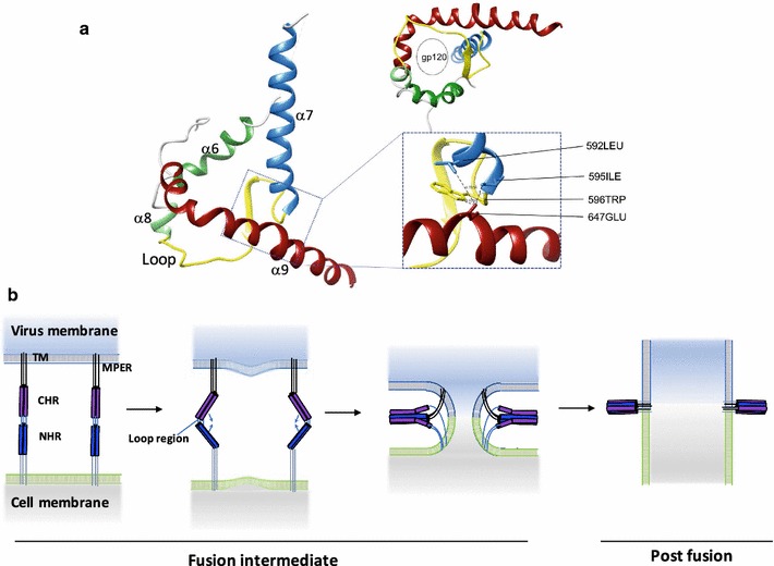
Background: The native pre-fusion structure of gp120/gp41 complex of human immunodeficiency virus type 1 was recently revealed. In the model, the helices of gp41 (α6, α7, α8, and α9) form a four-helix collar underneath trimeric gp120. Gp41 is a class I fusion protein and mediates membrane fusion by forming a post-fusion structure called the six-helix bundle (6HB). The comparison of the pre- and post-fusion structures revealed the large conformational changes in gp41 during the antiparallel packing of the N- and C-terminal heptad repeats (NHRs and CHRs) in membrane fusion. Several mutagenesis studies of gp41 performed in the past were interpreted based on 6HB, the only available structure at that time. To obtain an insight about the current pre-fusion structural model and conformational changes during membrane fusion, alanine insertion mutagenesis of the NHR, CHR and connecting loop regions of HXB2 gp41 was performed. The effects of mutations on biosynthesis and membrane fusion were analyzed by immunoblotting and fusion assays, respectively. The extent of membrane fusion was evaluated by split luciferase-based pore formation and syncytia formation assays, respectively.
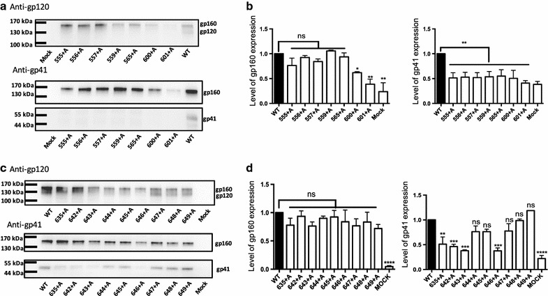
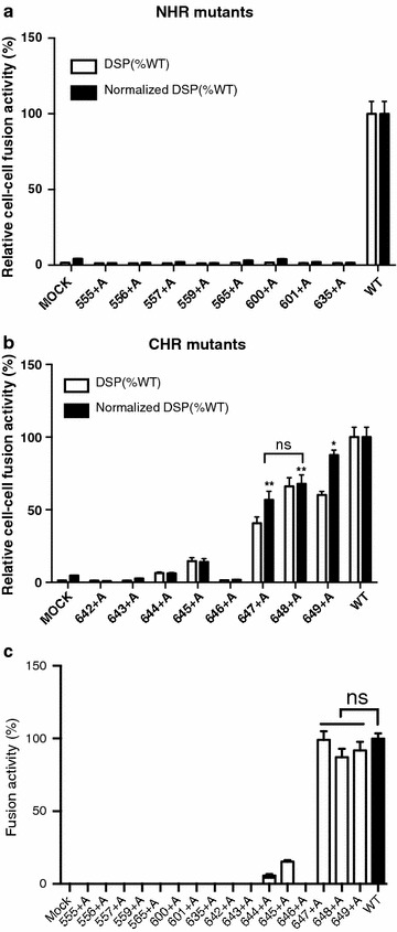
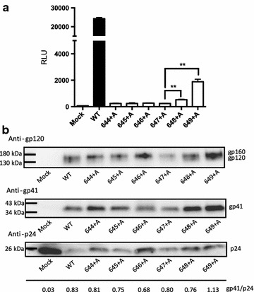
Results: Consistent with the current structural model, drastic negative effects of mutations on biosynthesis and membrane fusion were observed for NHR, loop, and proximal regions of CHR (up to amino acid position 643). The insertions in α9 after it leaves the four-helix collar were tolerable for biosynthesis. These CHR mutants showed varying effects on membrane fusion. Insertion at position 644 or 645 resulted in poor pore and syncytia formation. Efficient pore and syncytia formation almost similar to that of the wild type was observed for insertion at position 647, 648 or 649. However, recovery of virus infectivity was only observed for the insertions beyond position 648.
Conclusions: The mutagenesis data for HXB2 gp41 is in agreement with the recent pre-fusion structure model. The virus infection data suggested that fusion pores sufficiently large enough for the release of the virus genome complex are formed after the completion of 6HB beyond position 648.
Keywords: Envelope protein; Fusion pore; Heptad repeat; Human immunodeficiency virus type 1; Membrane fusion; Six-helix bundle; Split green fluorescent protein; Split luciferase.
Figures

Similar articles
-
Cell-cell and virus-cell fusion assay-based analyses of alanine insertion mutants in the distal α9 portion of the JRFL gp41 subunit from HIV-1.J Biol Chem. 2019 Apr 5;294(14):5677-5687. doi: 10.1074/jbc.RA118.004579. Epub 2019 Feb 8. J Biol Chem. 2019. PMID: 30737278 Free PMC article.
-
HIV-1 gp41 Residues Modulate CD4-Induced Conformational Changes in the Envelope Glycoprotein and Evolution of a Relaxed Conformation of gp120.J Virol. 2018 Jul 31;92(16):e00583-18. doi: 10.1128/JVI.00583-18. Print 2018 Aug 15. J Virol. 2018. PMID: 29875245 Free PMC article.
-
Conserved Residue Asn-145 in the C-Terminal Heptad Repeat Region of HIV-1 gp41 is Critical for Viral Fusion and Regulates the Antiviral Activity of Fusion Inhibitors.Viruses. 2019 Jul 3;11(7):609. doi: 10.3390/v11070609. Viruses. 2019. PMID: 31277353 Free PMC article.
-
Biochemistry and biophysics of HIV-1 gp41 - membrane interactions and implications for HIV-1 envelope protein mediated viral-cell fusion and fusion inhibitor design.Curr Top Med Chem. 2011 Dec;11(24):2959-84. doi: 10.2174/156802611798808497. Curr Top Med Chem. 2011. PMID: 22044229 Free PMC article. Review.
-
High throughput screening and characterization of HIV-1 entry inhibitors targeting gp41: theories and techniques.Curr Pharm Des. 2004;10(15):1827-43. doi: 10.2174/1381612043384466. Curr Pharm Des. 2004. PMID: 15180543 Review.
Cited by
-
Cell-cell and virus-cell fusion assay-based analyses of alanine insertion mutants in the distal α9 portion of the JRFL gp41 subunit from HIV-1.J Biol Chem. 2019 Apr 5;294(14):5677-5687. doi: 10.1074/jbc.RA118.004579. Epub 2019 Feb 8. J Biol Chem. 2019. PMID: 30737278 Free PMC article.
References
Publication types
MeSH terms
Substances
LinkOut - more resources
Full Text Sources
Other Literature Sources
Medical
Miscellaneous
