Review
. 2019 Feb 1;9(2):a031534.
doi: 10.1101/cshperspect.a031534.

The Interdependent Activation of Son-of-Sevenless and Ras

Affiliations
Review

The Interdependent Activation of Son-of-Sevenless and Ras

Pradeep Bandaru et al. Cold Spring Harb Perspect Med. .

Abstract

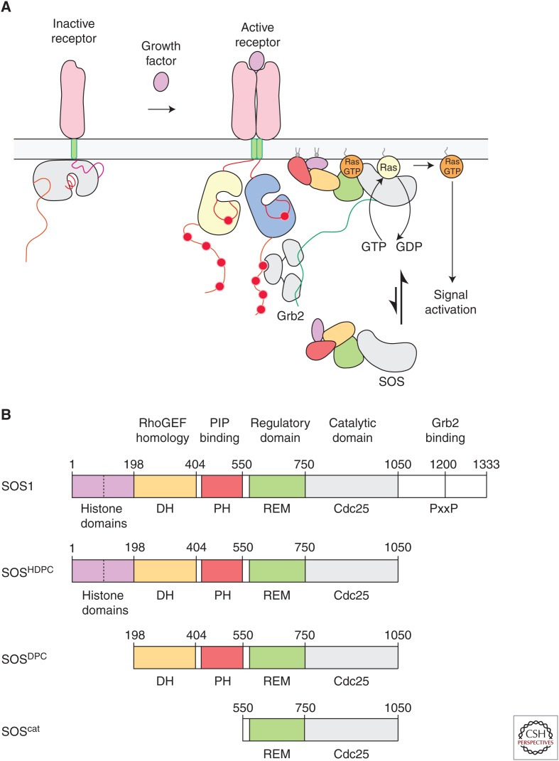
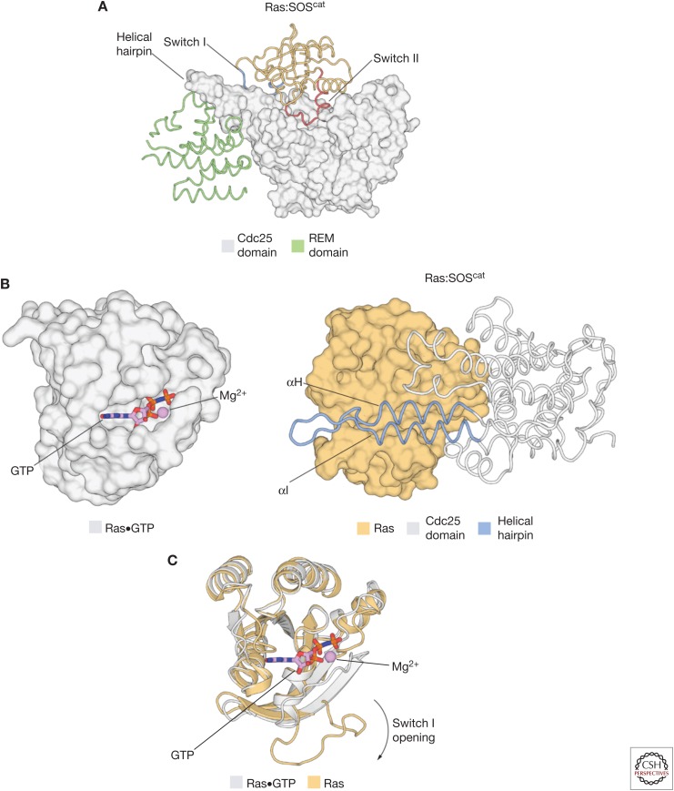
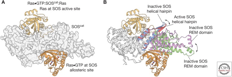
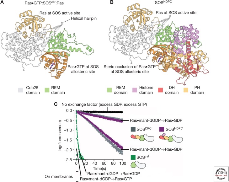
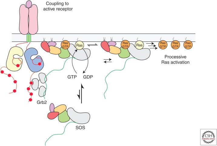
The guanine-nucleotide exchange factor (GEF) Son-of-Sevenless (SOS) plays a critical role in metazoan signaling by converting Ras•GDP (guanosine diphosphate) to Ras•GTP (guanosine triphosphate) in response to tyrosine kinase activation. Structural studies have shown that SOS differs from other Ras-specific GEFs in that SOS is itself activated by Ras•GTP binding to an allosteric site, distal to the site of nucleotide exchange. The activation of SOS involves membrane recruitment and conformational changes, triggered by lipid binding, that open the allosteric binding site for Ras•GTP. This is in contrast to other Ras-specific GEFs, which are activated by second messengers that more directly affect the active site. Allosteric Ras•GTP binding stabilizes SOS at the membrane, where it can turn over other Ras molecules processively, leading to an ultrasensitive response that is distinct from that of other Ras-specific GEFs.


Figures

Figure 1.
Figure 1.
The activation and domain organization of Son-of-Sevenless (SOS). (A) Ras activation is coupled to the phosphorylation of tyrosine residues on cell-surface receptors. This primarily occurs through the SH2-mediated recruitment of SOS to these receptors by adaptor proteins such as Grb2. Ras activation by SOS subsequently triggers downstream signaling responses in cells. (B) Schematic representation of the various constructs of SOS discussed in this review. DH, Dbl homology; PH, Pleckstrin homology; REM, Ras exchanger motif.
Figure 2.
Figure 2.
The structural mechanism of Ras activation by Son-of-Sevenless (SOS). (A) The structure of the Ras:SOScat complex (Protein Data Bank [PDB] code: 1BKD) showed that SOS stabilizes the nucleotide-free form of Ras by forming an extensive interface involving Switch I and Switch II of Ras. (B) (Left) The structure of Ras•GTP (PDB code: 5P21). (Right) A helical hairpin element that protrudes from the main body of the Cdc25 domain of SOS is inserted between the Switch I and Switch II elements of Ras, and causes the active site of Ras to open widely and expel the nucleotide. (C) After the insertion of the helical hairpin of SOS into Ras, the principal conformational change in Ras that leads to nucleotide release is a peeling away of Switch I from the rest of the protein. REM, Ras exchanger motif; GTP, guanosine triphosphate.
Figure 3.
Figure 3.
The activation of Son-of-Sevenless (SOS) by Ras•GTP (guanosine triphosphate). (A) The structure of the 2:1 Ras:SOScat complex (Protein Data Bank [PDB] code: 1NVV) revealed that nucleotide-free Ras was bound to the active site of the Cdc25 domain, and that a second Ras•GTP molecule is bound to a distal allosteric site, wedged between the Ras exchanger motif (REM) and Cdc25 domains (B) Comparison of the Ras-free structure of SOScat (PDB code: 2II0) to the 2:1 Ras:SOScat complex shows that the helical hairpin is tilted toward the active site of SOS in the Ras-free state, thereby constricting the site where SOS engages Switch II of nucleotide-free Ras. Allosteric binding of Ras•GTP to SOS leads to the rotation and opening of the helical hairpin, accompanied by the outward rotation of the REM domain, which frees the catalytic site to bind Ras.
Figure 4.
Figure 4.
The autoinhibition of Son-of-Sevenless (SOS) by regulatory domains. (A) The structure of the 2:1 Ras:SOScat complex, depicting Ras bound to the Cdc25 domain and Ras•GTP (guanosine triphosphate) bound to the allosteric site of SOS. (B) The structure of SOS containing the Dbl homology (DH)-Pleckstrin homology (PH) module and the catalytic module (SOSDPC) (Protein Data Bank [PDB] code: 3KSY) show that the DH-PH module sterically occludes the binding of Ras•GTP at the allosteric site. (C) Biochemical measurements show that SOSDPC has lower activity than SOScat. The fastest traces are for membrane-bound Ras (Gureasko et al. 2008). REM, Ras exchanger motif; GDP, guanosine diphosphate; dGDP, deoxyguanosine diphosphate.
Figure 5.
Figure 5.
The activation of Son-of-Sevenless (SOS) at the membrane. SOS is initially recruited to the membrane by adaptor proteins, such as Grb2, that bind to activated cell-surface receptors. Once a single molecule of SOS is activated allosterically by Ras•GTP (guanosine triphosphate) at the membrane, many more Ras molecules can then be activated processively by that SOS molecule, even if it were to disengage from the receptor. GDP, Guanosine diphosphate.

References

    1. Aghazadeh B, Lowry WE, Huang XY, Rosen MK. 2000. Structural basis for relief of autoinhibition of the Dbl homology domain of proto-oncogene Vav by tyrosine phosphorylation. Cell 102: 625–633. - PubMed
    1. Aronheim A, Engelberg D, Li N, al-Alawi N, Schlessinger J, Karin M. 1994. Membrane targeting of the nucleotide exchange factor Sos is sufficient for activating the Ras signaling pathway. Cell 78: 949–961. - PubMed
    1. Bandaru P, Shah NH, Bhattacharyya M, Barton JP, Kondo Y, Cofsky JC, Gee CL, Chakraborty AK, Kortemme T, Ranganathan R, Kuriyan, J. 2017. Deconstruction of the Ras switching cycle through saturation mutagenesis. eLife 6: e27810. - PMC - PubMed
    1. Baxevanis AD, Arents G, Moudrianakis EN, Landsman D. 1995. A variety of DNA-binding and multimeric proteins contain the histone fold motif. Nucleic Acids Res 23: 2685–2691. - PMC - PubMed
    1. Bennett BD, Kimball EH, Gao M, Osterhout R, Van Dien SJ, Rabinowitz JD. 2009. Absolute metabolite concentrations and implied enzyme active site occupancy in Escherichia coli. Nat Chem Biol 5: 593–599. - PMC - PubMed

Publication types