. 2018 Apr 17;115(16):4270-4275.
doi: 10.1073/pnas.1716479115. Epub 2018 Apr 2.

Ectopic neurogenesis induced by prenatal antiepileptic drug exposure augments seizure susceptibility in adult mice

Affiliations

Ectopic neurogenesis induced by prenatal antiepileptic drug exposure augments seizure susceptibility in adult mice

Atsuhiko Sakai et al. Proc Natl Acad Sci U S A. .

Abstract

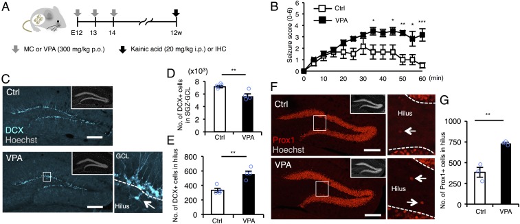
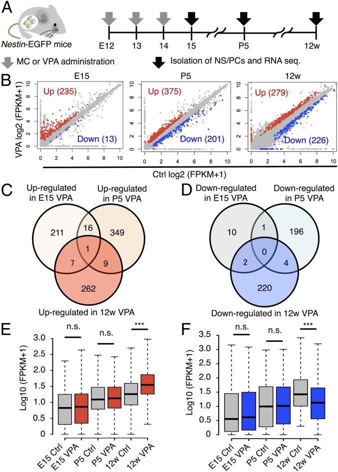
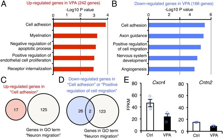
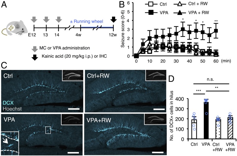
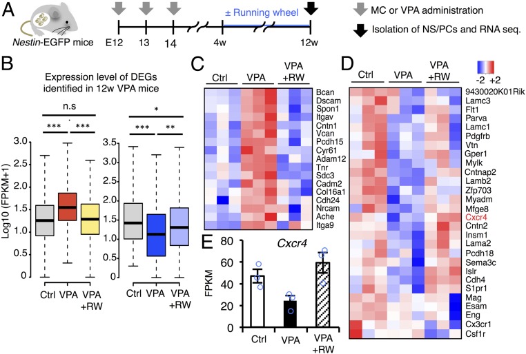
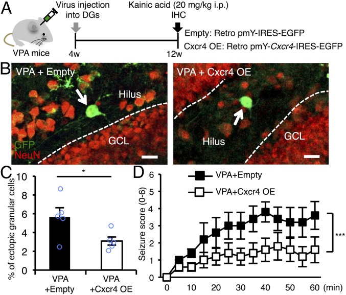
Epilepsy is a neurological disorder often associated with seizure that affects ∼0.7% of pregnant women. During pregnancy, most epileptic patients are prescribed antiepileptic drugs (AEDs) such as valproic acid (VPA) to control seizure activity. Here, we show that prenatal exposure to VPA in mice increases seizure susceptibility in adult offspring through mislocalization of newborn neurons in the hippocampus. We confirmed that neurons newly generated from neural stem/progenitor cells (NS/PCs) are integrated into the granular cell layer in the adult hippocampus; however, prenatal VPA treatment altered the expression in NS/PCs of genes associated with cell migration, including CXC motif chemokine receptor 4 (Cxcr4), consequently increasing the ectopic localization of newborn neurons in the hilus. We also found that voluntary exercise in a running wheel suppressed this ectopic neurogenesis and countered the enhanced seizure susceptibility caused by prenatal VPA exposure, probably by normalizing the VPA-disrupted expression of multiple genes including Cxcr4 in adult NS/PCs. Replenishing Cxcr4 expression alone in NS/PCs was sufficient to overcome the aberrant migration of newborn neurons and increased seizure susceptibility in VPA-exposed mice. Thus, prenatal exposure to an AED, VPA, has a long-term effect on the behavior of NS/PCs in offspring, but this effect can be counteracted by a simple physical activity. Our findings offer a step to developing strategies for managing detrimental effects in offspring exposed to VPA in utero.

Keywords: Cxcr4; ectopic neurogenesis; epilepsy; neural stem cell; valproic acid.


Conflict of interest statement

The authors declare no conflict of interest.

Figures

Fig. 1.
Fig. 1.
Prenatal exposure to VPA increases seizure susceptibility and ectopic hippocampal neurogenesis in the adult. (A) Experimental scheme for investigating seizure susceptibility and adult neurogenesis. Control [administered with methylcellulose (MC)] and VPA mice were randomly assigned to the groups for scoring of seizure severity and immunohistochemistry (IHC). (B) Seizure response to KA treatment over time in control and VPA mice (n = 6 animals each). Two-way repeated measures ANOVA was used for statistical analysis (treatment: F1,130 = 59.08, P < 0.0001; time: F12,130 = 7.063, P < 0.0001; treatment × time interaction: F12,130 = 2.341, P = 0.0095, post hoc Bonferroni’s multiple comparison test). (C) Representative images of DCX-positive (cyan) immature neurons in the DG. The area outlined by a white rectangle in the lower main panel is enlarged to the right. The arrow indicates a DCX-labeled cell in the hilus, and dashed white lines mark the boundaries between GCL and hilus. (Insets) H33258 nuclear staining of each field. (Scale bar, 200 µm.) (D and E) Quantification of the number of DCX-positive cells in the SGZ/GCL (D) and hilus (E) (n = 4 animals each). (F) Representative images of Prox1-positive (red) GCs in the DG. The area outlined by a white rectangle in the lower main panel is enlarged to the right. The arrow indicates a Prox1-positive cell in the hilus, and the dashed white line marks the boundary between GCL and hilus. (Insets) H33258 nuclear staining (gray) of each field. (Scale bar, 200 µm.) (G) Quantification of the number of Prox1-positive cells in the hilus (n = 3 animals each). *P ≤ 0.05, **P ≤ 0.01, ***P ≤ 0.001.
Fig. 2.
Fig. 2.
Transcriptome analysis of NS/PCs from control and VPA mice at distinct developmental stages. (A) Schematic representation of the isolation of NS/PCs from Ctrl and VPA mice at E15, P5, and 12w. RNAs were extracted from these cells and subjected to sequence analysis. (B) Scatter plots of genes expressed in NS/PCs from Ctrl and VPA mice. Up- (red) and down-regulated (blue) DEGs are highlighted. (C and D) Venn diagrams of up- (C) and down-regulated (D) DEGs at each developmental stage. (E and F) Box plot of up- (E) and down-regulated (F) DEG expression in NS/PCs of 12w hippocampal DG. In contrast to 12w, expression levels of the DEGs are comparable between Ctrl and VPA mice at E15 and P5. ***P ≤ 0.001. n.s., not significant.
Fig. 3.
Fig. 3.
Prenatal VPA exposure alters the expression level of cell migration-related genes in NS/PCs of adult DG. (A and B) Functional annotation of up- (A) and down-regulated (B) genes in NS/PCs of adult VPA mice relative to control mice. The top five GO terms in each gene group are displayed. (C) Venn diagram of up-regulated genes categorized in the GO term “cell adhesion” and the gene list associated with “neuron migration.” There was no overlap between genes in each category. (D) Identification of two candidate genes for the ectopic neuronal migration in VPA mice at 12w. Down-regulated genes categorized in the GO terms “cell adhesion” or “positive regulation of cell migration” overlapped with two genes in the GO term “neuron migration.” (E) Expression levels of the two genes identified in D.
Fig. 4.
Fig. 4.
Voluntary exercise alleviates increased seizure susceptibility and abnormal neuronal migration in VPA mice. (A) Experimental scheme for investigating the effect of voluntary running on seizure susceptibility and neuronal migration in the DG. Control (administered with MC) and VPA mice were randomly assigned to the groups for scoring of seizure severity and IHC. (B) Seizure response to KA treatment over time in control and VPA mice with or without voluntary running as indicated (n = 6 animals each). Two-way repeated measures ANOVA was used for statistical analysis (treatment: F3,240 = 3.052, P = 0.0522; time: F12,240 = 9.837, P < 0.0001; treatment × time interaction: F36,240 = 2.428, P < 0.0001, post hoc Bonferroni’s multiple comparison test for VPA vs. VPA+RW, *P ≤ 0.05, **P ≤ 0.01). (C) Representative images of DCX-positive immature neurons (cyan) in the DG. The area outlined by the white rectangle in the lower left is magnified in the inset. The arrow indicates a DCX-positive cell in the hilus, and the dashed white line marks the boundary between hilus and GCL. (Insets) H33258 nuclear staining (gray) of each field. (Scale bar, 200 µm.) (D) Quantification of the number of DCX-positive cells in the hilus (n = 5 animals each). One-way ANOVA was used for statistical analysis (F3,16 = 13.31, P < 0.0001, post hoc Tukey’s multiple comparison test, **P ≤ 0.01, ***P ≤ 0.001. n.s., not significant).
Fig. 5.
Fig. 5.
Voluntary running normalizes transcriptomic alteration of adult NS/PCs in VPA mice. (A) Schematic representation of the isolation of NS/PCs from Ctrl and VPA mice at 12w. RNAs were extracted from these cells and subjected to sequence analysis. (B) Box plots of up- (Left) and down-regulated (Right) DEG expression in NS/PCs of 12w hippocampal DG. Changes of gene expression, in both directions, after VPA exposure were largely normalized by running (RW); *P ≤ 0.05, **P ≤ 0.01, ***P ≤ 0.001, n.s indicates not significant. (C) Heat map showing the expression level of up-regulated DEGs in adult VPA mice categorized in the GO term “cell adhesion” (Fig. 2 B and D) in control, VPA, and VPA+RW mice. (D) Heat map indicating the expression level of down-regulated genes in the adult VPA mice categorized in GO term “cell adhesion” and “positive regulation of cell migration” (Fig. 2 C and E) in control, VPA, and VPA+RW mice. (E) Expression level of Cxcr4 in NS/PCs of 12w control, VPA, and VPA+RW mice.
Fig. 6.
Fig. 6.
Replenishment of Cxcr4 expression in NS/PCs of the DG alleviates the increased seizure susceptibility and abnormal neuronal migration in VPA mice. (A) Experimental scheme for investigating the effect of Cxcr4 expression in NS/PCs on seizure susceptibility and neuronal migration in VPA mice. (B) Representative images of GFP (green) and NeuN (red) dual-positive (GFP+NeuN+) newborn neurons located in the hilus (arrows). Dashed white lines indicate the boundary between hilus and GCL. (Scale bar, 20 µm.) (C) Quantification of percentages of the number of ectopically located GFP+NeuN+ cells among total GFP+NeuN+ cells in the DG (n = 5 animals each). (D) Seizure response to KA treatment over time in VPA mice that received control and Cxcr4-expressing retrovirus injection (n = 5 animals each). Two-way repeated measures ANOVA was used for statistical analysis (Cxcr4: F1,104 = 33.14, P < 0.0001; time: F12,104 = 3.344, P = 0.0004; Cxcr4 × time interaction: F12,104 = 0.5549, P = 0.8731). *P ≤ 0.05, ***P ≤ 0.001.

Comment in

References

    1. Tomson T, et al. EURAP study group Dose-dependent risk of malformations with antiepileptic drugs: An analysis of data from the EURAP epilepsy and pregnancy registry. Lancet Neurol. 2011;10:609–617. - PubMed
    1. Dean JC, et al. Long term health and neurodevelopment in children exposed to antiepileptic drugs before birth. J Med Genet. 2002;39:251–259. - PMC - PubMed
    1. Viinikainen K, Heinonen S, Eriksson K, Kälviäinen R. Community-based, prospective, controlled study of obstetric and neonatal outcome of 179 pregnancies in women with epilepsy. Epilepsia. 2006;47:186–192. - PubMed
    1. Jentink J, et al. EUROCAT Antiepileptic Study Working Group Valproic acid monotherapy in pregnancy and major congenital malformations. N Engl J Med. 2010;362:2185–2193. - PubMed
    1. Meador KJ, et al. NEAD Study Group Cognitive function at 3 years of age after fetal exposure to antiepileptic drugs. N Engl J Med. 2009;360:1597–1605. - PMC - PubMed

Publication types

MeSH terms