. 2018 Jan 30;5(4):198-207.
doi: 10.15698/mic2018.04.625.

Impact of F1Fo-ATP-synthase dimer assembly factors on mitochondrial function and organismic aging

Affiliations

Impact of F1Fo-ATP-synthase dimer assembly factors on mitochondrial function and organismic aging

Nadia G Rampello et al. Microb Cell. .

Abstract

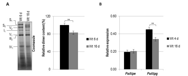
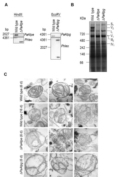
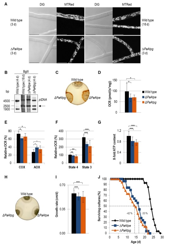
In aerobic organisms, mitochondrial F1Fo-ATP-synthase is the major site of ATP production. Beside this fundamental role, the protein complex is involved in shaping and maintenance of cristae. Previous electron microscopic studies identified the dissociation of F1Fo-ATP-synthase dimers and oligomers during organismic aging correlating with a massive remodeling of the mitochondrial inner membrane. Here we report results aimed to experimentally proof this impact and to obtain further insights into the control of these processes. We focused on the role of the two dimer assembly factors PaATPE and PaATPG of the aging model Podospora anserina. Ablation of either protein strongly affects mitochondrial function and leads to an accumulation of senescence markers demonstrating that the inhibition of dimer formation negatively influences vital functions and accelerates organismic aging. Our data validate a model that links mitochondrial membrane remodeling to aging and identify specific molecular components triggering this process.

Keywords: F1Fo-ATP-synthase; aging; membranes; mitochondria; remodeling.


Conflict of interest statement

Conflict of interest: The authors declare that they have no conflict of interest.

Figures

Figure 1
Figure 1. FIGURE 1: Age-dependent dimer decrease and analysis of expression of PaAtpe and PaAtpg in P. anserina.
(A) Representative BN-PAGE analysis and quantification of mitochondrial protein extracts (V2, F1Fo-ATP-synthase dimers) from three independent 6-days and 16-days old wild-type strains. F1Fo-ATP-synthase dimers were normalized to Coomassie stained gel. Relative dimer content of 6-days old wild-type strains was set to 100%. Mitochondrial protein complexes were stained with Coomassie Blue and complexes and supercomplexes S1, S0, V2, I1, V1, III1 and IV1 are indicated. (B) Transcript analysis of PaAtpe and PaAtpg in three young (4 d) and old (16 d) wild type cultures, respectively. RNA was isolated and gene expression was determined by qRT-PCR analyses. The relative expression was normalized to the expression of PaPorin coding for a mitochondrial outer membrane protein. Error bars represent the standard deviation and the P-values were determined by Student´s t test.
Figure 2
Figure 2. FIGURE 2: Deletion of PaAtpe or PaAtpg affects F1Fo-ATP-synthase dimer formation and mitochondrial inner membrane ultrastructure.
(A) Southern blot analysis of HindIII- and EcoRV-digested genomic DNA (gDNA) of wild-type, ΔPaAtpe and ΔPaAtpg strains. The digested gDNA was hybridized with a specific PaAtpe, PaAtpg and Phleo probe, respectively, and demonstrated a successful deletion of PaAtpe and PaAtpg. (B) Representative BN-PAGE analysis of mitochondrial protein extracts from 6-days old wild-type, ΔPaAtpe and ΔPaAtpg strains. Mitochondrial complexes including S1, V2, I1, V1, III1 and IV1 were visualized by Coomassie staining. (C) EM of isolated mitochondria of wild-type and deletion strains of different age (6 days and 16 days old). The morphology of at least 50 organelles per strain and growth condition was analyzed. Representative images are shown at the same magnification. Scale bar: 0,2 µm.
Figure 3
Figure 3. FIGURE 3: Impairments of mitochondrial functions affect growth and lifespan in deletion mutants.
(A) Comparison of mitochondrial morphology of 3-days and 16-days old wild-type strains with those of ΔPaAtpe and ΔPaAtpg (3 d) by fluorescence microscopy. Mitochondria were stained with MitoTracker® Red and analyzed by confocal fluorescence microscopy. Scale bars: 10 µm. (B) Representative Southern blot analysis of BglII-digested gDNA of wild-type (4-days and 16-days old), ΔPaAtpe and ΔPaAtpg strains (4-days old). The 2500 bp age-specific plDNA (arrow) fragment was detected with a specific plDNA probe. (C) Release of ROS species H2O2 from wild-type (6-days n=3), ΔPaAtpe (6-days n=3) and ΔPaAtpg strains (6-days n = 3) visualized by DAB precipitation. (D) Oxygen consumption (basal respiration) of 6-days old deletion mutant mycelium compared to respiration of 6-days old wild type mycelium (n = 3-4 biological replicates with a total number of 12-16 technical replicates). (E) Relative COX- and AOX-dependent oxygen consumption rate (OCR) of 6-days old wild-type and 6-days old deletion strains mycelium after treatment with KCN (COX-inhibitor) or SHAM (AOX-inhibitor). (F) Relative complex I-dependent OCR of 6-days old wild-type mitochondria compared to mitochondria from 6-days old deletion mutants (n = 4-8 biological replicates with a total number of 15-28 technical replicates). State 4 OCR of mitochondria from wild type was set to 100%. (G) ATP production of 6-days old wild-type and 6-days old deletion mutant mitochondria after removal of samples during state 3 respiration measurements. Samples of three biological replicates and two technical replicates, respectively, were measured three times by a luminescence-based assay. (H) Phenotypic analysis of indicated strains growing on M2 medium for 4 days. (I) Growth rate of wild type (0.58 ± 0.05 cm/d, n = 27), ΔPaAtpe (0.53 ± 0.05 cm/d, n = 25) and ΔPaAtpg (0.53 ± 0.08 cm/d, n =3 3) growing on M2 medium. Growth rate was determined from day 3 to 6 of growth. (J) Lifespan analysis of wild type (mean lifespan = 23.3 d, n = 27), ΔPaATPe (mean lifespan = 15.1 d, n = 25,) and ΔPaATPg (mean lifespan = 13.5 d, n = 33) growing on M2 medium. The error bars represent the standard deviation and the P-values were determined by Student´s t test.

References

    1. Zick M, Rabl R, Reichert AS. Cristae formation-linking ultrastructure and function of mitochondria. Biochim Biophys Acta. 2009;1793(1):5–19. doi: 10.1016/j.bbamcr.2008.06.013. - DOI - PubMed
    1. Cogliati S, Enriquez JA, Scorrano L. Mitochondrial cristae: where beauty meets functionality. Trends Biochem Sci. 2016;41(3):261–273. doi: 10.1016/j.tibs.2016.01.001. - DOI - PubMed
    1. Harner ME, Unger AK, Geerts WJ, Mari M, Izawa T, Stenger M, Geimer S, Reggiori F, Westermann B, Neupert W. An evidence based hypothesis on the existence of two pathways of mitochondrial crista formation. Elife. 2016;5:e18853. doi: 10.7554/eLife.18853. - DOI - PMC - PubMed
    1. Rampelt H, Zerbes RM, van der Laan M, Pfanner N. Role of the mitochondrial contact site and cristae organizing system in membrane architecture and dynamics. Biochim Biophys Acta. 2017;1864(4):737–746. doi: 10.1016/j.bbamcr.2016.05.020. - DOI - PubMed
    1. Jonckheere AI, Smeitink JA, Rodenburg RJ. Mitochondrial ATP synthase: architecture, function and pathology. J Inherit Metab Dis. 2012;35(2):211–225. doi: 10.1007/s10545-011-9382-9. - DOI - PMC - PubMed

LinkOut - more resources