. 2017 Dec 1;49(12):e405.
doi: 10.1038/emm.2017.200.

Intracellular trafficking of TREM2 is regulated by presenilin 1

Affiliations

Intracellular trafficking of TREM2 is regulated by presenilin 1

Yingjun Zhao et al. Exp Mol Med. .

Abstract

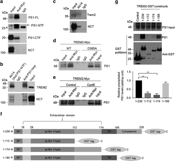
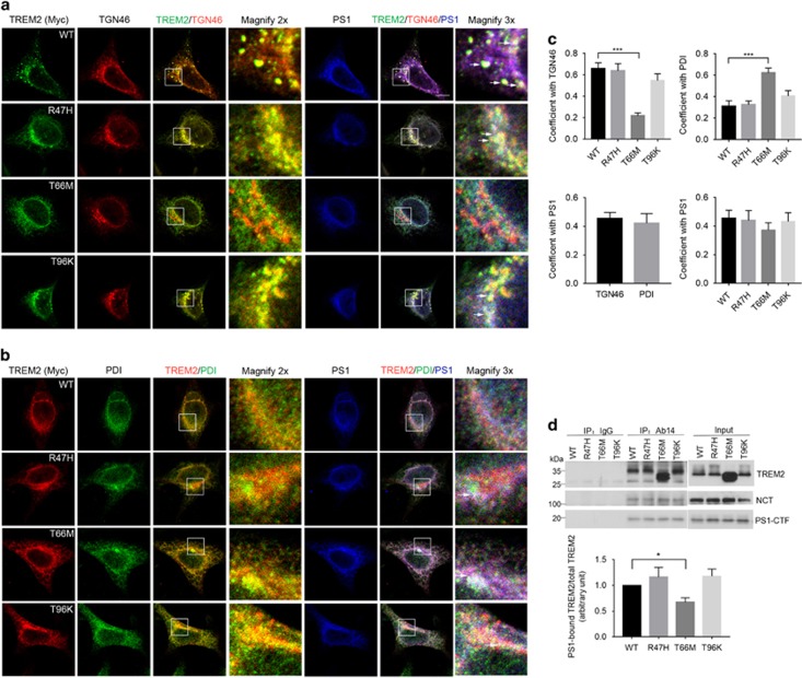
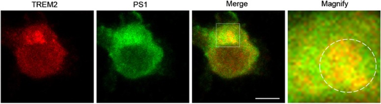
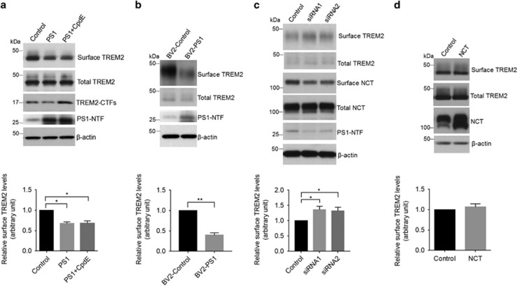
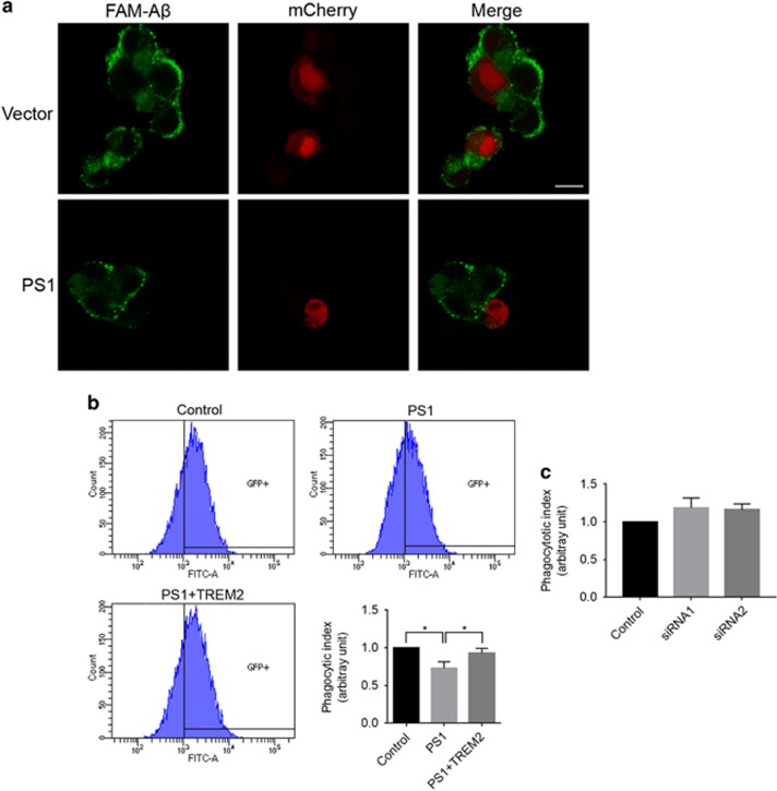
Genetic mutations in triggering receptor expressed on myeloid cells 2 (TREM2) have been linked to a variety of neurodegenerative diseases including Alzheimer's disease, amyotrophic lateral sclerosis, frontotemporal dementia and Parkinson's disease. In the brain, TREM2 is highly expressed on the cell surface of microglia, where it can transduce signals to regulate microglial functions such as phagocytosis. To date, mechanisms underlying intracellular trafficking of TREM2 remain elusive. Mutations in the presenilin 1 (PS1) catalytic subunit of the γ-secretase complex have been associated with increased generation of the amyloidogenic Aβ (amyloid-β) 42 peptide through cleavage of the Aβ precursor amyloid precursor protein. Here we found that TREM2 interacts with PS1 in a manner independent of γ-secretase activity. Mutations in TREM2 alter its subcellular localization and affects its interaction with PS1. Upregulation of PS1 reduces, whereas downregulation of PS1 increases, steady-state levels of cell surface TREM2. Furthermore, PS1 overexpression results in attenuated phagocytic uptake of Aβ by microglia, which is reversed by TREM2 overexpression. Our data indicate a novel role for PS1 in regulating TREM2 intracellular trafficking and pathophysiological function.


Conflict of interest statement

The authors declare no conflict of interest.

Figures

Figure 1
Figure 1
Presenilin 1 (PS1) interacts with TREM2. (a, b) PS1 constructs were transfected into HEK293 cells stably expressing TREM2 with a Myc-tag at the C terminus (HEK293-TREM2). (a) Cell lysates were immunoprecipitated with a Myc antibody or control IgG. Immunoprecipitated proteins were subjected to immunoblotting with an Ab14 antibody to detect full-length PS1 (PS1-FL) and the PS1 N-terminal fragment (NTF), an anti-PS1 loop antibody to detect the PS1 C-terminal fragment (CTF) and a nicastrin (NCT) antibody as indicated. (b) Cell lysates were immunoprecipitated with Ab14, anti-PS1 loop or control IgG and immunoblotted with Myc and NCT antibodies. (c) Lysates from BV2 microglial cells were immunoprecipitated with Ab14 and immunoblotted with NCT and mouse TREM2 antibodies. (d) Vectors expressing wild-type (WT) or mutant PS1 (D385A) were transfected into HEK293-TREM2 cells. Cell lysates were immunoprecipitated with Ab14 or control IgG, and PS1 was detected by immunoblotting. (e) Lysates from HEK293-TREM2 cells with or without Compound E (CpdE, a γ-secretase inhibitor) treatment were immunoprecipitated with Ab14 or control IgG, and PS1 was detected by immunoblotting. (f) Schematic representations of full-length (1–230) or truncated TREM2 constructs, all tagged with GST at the C terminus. SP, signal peptide; TM, transmembrane domain. (g) PS1 was co-expressed with full-length TREM2 or other TREM2 fragments as shown in f in HEK293 cells. Cell lysates were precipitated with Glutathione Sepharose beads and immunoblotted with the PS1 antibody Ab14 or an antibody against GST. PS1 co-precipitation levels were determined by densitometric analysis and normalized with respect to both PS1 expression and precipitated GST. **P<0.01, n=3, Student’s t-test.
Figure 2
Figure 2
Mutations in TREM2 affect colocalization and interactions between TREM2 and presenilin 1 (PS1). PS1 was transfected into HEK293 cells stably expressing Myc-tagged TREM2 WT or TREM2 mutants as indicated. (a, b) Cells were then subjected to immunostaining with antibodies against Myc, PS1, and TGN46 (a marker for the Golgi, a) or PDI (a marker for the ER, b). White arrows in magnified images indicate colocalizing overlap for TREM2, PS1 and TGN46/PDI. Scale bars for a, b, 10 μm. (c) Quantification of colocalized signals. Pearson’s correlation coefficient is shown. ***P<0.001, n=3 independent experiments, one-way ANOVA with Dunnett’s post hoc analysis. (d) Cell lysates were immunoprecipitated with the PS1 antibody Ab14 or normal IgG. TREM2, NCT and PS1-CTF were detected by immunoblotting. The levels of precipitated TREM2 WT and mutants were normalized to the input. *P<0.05, n=3, Student’s t-test.
Figure 3
Figure 3
Endogenous TREM2 partially colocalizes with endogenous presenilin 1 (PS1) in microglial BV2 cells. BV2 cells were immunostained with antibodies against PS1 and mouse TREM2. White circles in magnified images indicate some colocalizing overlap between TREM2 and PS1 in Golgi-like structures. Scale bar, 5 μm.
Figure 4
Figure 4
Upregulation of presenilin 1 (PS1) reduces steady-state levels of TREM2 at the cell surface. (a) Following PS1 overexpression, HEK293-TREM2 cells were treated with or without the γ-secretase inhibitor Compound E (CpdE) and subjected to cell surface biotinylation assay. Precipitates from streptavidin-agarose beads were immunoblotted for biotinylated TREM2 and total TREM2 levels (levels of TREM2 in 2% total cell lysates). *P<0.05, n=3, one-way ANOVA with Sidak post hoc analysis. (b) The level of endogenous TREM2 at the surface of microglial BV2 cells stably expressing PS1 (BV2-PS1) was determined by biotinylation. **P<0.01, n=3, Student’s t-test. (c) BV2 cells were transfected with a scrambled control siRNA or PS1-targeting siRNAs for 72 h. The level of cell surface TREM2 was determined by surface biotinylation. *P<0.05, n=3, Student’s t-test. (d) Cell surface expression of TREM2 in HEK293-TREM2 cells with NCT overexpression, as determined by biotinylation. n=3, Student’s t-test.
Figure 5
Figure 5
Overexpression of presenilin 1 (PS1) impairs TREM2-mediated phagocytosis in microglial cells. (a) PS1 and mCherry were co-transfected into BV2 microglial cells. The cells were then incubated with 6-carboxyfluorescein (FAM)-labeled Aβ42 for 2 h. FAM-Aβ42 uptake was analyzed by fluorescence microscopy. Scale bars, 10 μm. (b) Phagocytosis of FAM-Aβ42 in BV2 cells stably expressing PS1 as determined by flow cytometry. *P<0.05, n=3, one-way ANOVA with Sidak post hoc analysis. (c) Phagocytosis of FAM-Aβ42 in BV2 cells following PS1 knockdown as determined by flow cytometry. n=3, Student’s t-test.

References

    1. Guerreiro RJ, Lohmann E, Bras JM, Gibbs JR, Rohrer JD, Gurunlian N et al. Using exome sequencing to reveal mutations in TREM2 presenting as a frontotemporal dementia-like syndrome without bone involvement. JAMA Neurol 2013; 70: 78–84. - PMC - PubMed
    1. Giraldo M, Lopera F, Siniard AL, Corneveaux JJ, Schrauwen I, Carvajal J et al. Variants in triggering receptor expressed on myeloid cells 2 are associated with both behavioral variant frontotemporal lobar degeneration and Alzheimer's disease. Neurobiol Aging 2013; 34: 2077 e11–2077 e18. - PMC - PubMed
    1. Rayaprolu S, Mullen B, Baker M, Lynch T, Finger E, Seeley WW et al. TREM2 in neurodegeneration: evidence for association of the p.R47H variant with frontotemporal dementia and Parkinson's disease. Mol Neurodegener 2013; 8: 19. - PMC - PubMed
    1. Le Ber I, De Septenville A, Guerreiro R, Bras J, Camuzat A, Caroppo P et al. Homozygous TREM2 mutation in a family with atypical frontotemporal dementia. Neurobiol Aging 2014; 35: 2419.e23–2419.e25. - PMC - PubMed
    1. Borroni B, Ferrari F, Galimberti D, Nacmias B, Barone C, Bagnoli S et al. Heterozygous TREM2 mutations in frontotemporal dementia. Neurobiol Aging 2014; 35: 934 e7–934.e10. - PubMed

Publication types

MeSH terms

LinkOut - more resources