

Molecular signatures of epithelial oviduct cells of a laying hen (Gallus gallus domesticus) and quail (Coturnix japonica)
- PMID: 29614966
- PMCID: PMC5883888
- DOI: 10.1186/s12861-018-0168-2
Molecular signatures of epithelial oviduct cells of a laying hen (Gallus gallus domesticus) and quail (Coturnix japonica)
Abstract
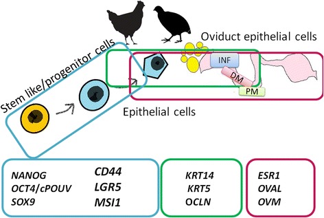
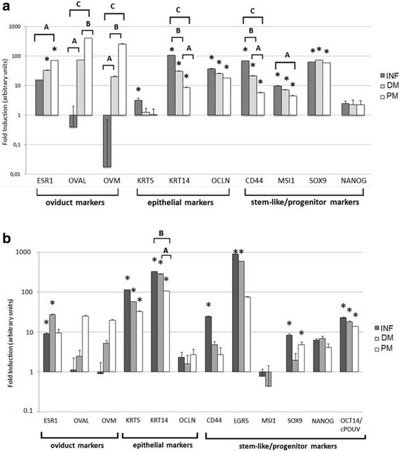
Background: In this work we have determined molecular signatures of oviduct epithelial and progenitor cells. We have proposed a panel of selected marker genes, which correspond with the phenotype of oviduct cells of a laying hen (Gallus gallus domesticus) and quail (Coturnix japonica). We demonstrated differences in characteristics of those cells, in tissue and in vitro, with respect to different anatomical and functional parts of the oviduct (infundibulum (INF), distal magnum (DM, and proximal magnum (PM)). The following gene expression signatures were studied: (1) oviduct markers (estrogen receptor 1, ovalbumin, and SPINK7 - ovomucoid), (2) epithelial markers (keratin 5, keratin 14, and occludin) and (3) stem-like/progenitor markers (CD44 glycoprotein, LGR5, Musashi-1, and sex determining region Y-box 9, Nanog homebox, OCT4/cPOUV gene encoding transcription factor POU5F3).
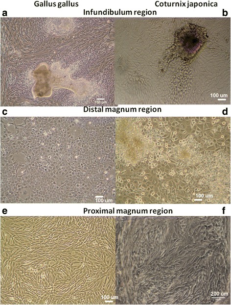
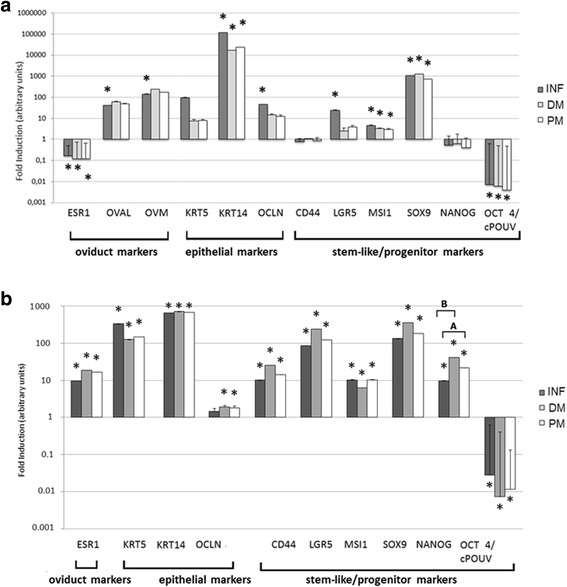
Results: In chicken, the expression of oviduct markers increased toward the proximal oviduct. Epithelial markers keratin14 and occludin were high in distal oviduct and decreased toward the proximal magnum. In quail oviduct tissue, the gene expression pattern of oviduct/epithelial markers was similar to chicken. The markers of progenitors/stemness in hen oviduct (Musashi-1 and CD44 glycoprotein) had the highest relative expression in the infundibulum and decreased toward the proximal magnum. In quail, we found significant expression of four progenitor markers (LGR5 gene, SRY sex determining region Y-box 9, OCT4/cPOUV gene, and CD44 glycoprotein) that were largely present in the distal oviduct. After in vitro culture of oviduct cells, the gene expression pattern has changed. High secretive potential of magnum-derived cells diminished by using decreased abundance of mRNA. On the other hand, chicken oviduct cells originating from the infundibulum gained ability to express OVM and OVAL. Epithelial character of the cells was maintained in vitro. Among progenitor markers, both hen and quail cells expressed high level of SOX9, LGR5 and Musashi-1.
Conclusion: Analysis of tissue material revealed gradual increase/decrease pattern in majority of the oviduct markers in both species. This pattern changed after the oviductal cells have been cultured in vitro. The results can provide molecular tools to validate the phenotype of in vitro biological models from reproductive tissue.
Keywords: Epithelial cells; Laying hen; Laying quail; Molecular signatures; Oviduct; Progenitor cells.
Conflict of interest statement
Ethics approval and consent to participate
The study was approved by the Local Ethics Committee for Animal Research (
Consent for publication
Not applicable.
Competing interests
The authors declare that they have no competing interests.
Publisher’s Note
Springer Nature remains neutral with regard to jurisdictional claims in published maps and institutional affiliations.
Figures
References
-
- Trevino LS, Johnson PA. Estrogen receptor subtype expression is altered in the hen model of ovarian Cancer. J Mol Genet Med 2016;10. 10.4172/1747-0862.1000203.
Publication types
MeSH terms
Substances
Grants and funding
- UMO-2011/03/N/NZ9/03814/National Science Centre Poland/International
- POLONIUM 2015-2017 granted to Professor Marek Bednarczyk and Dr Bertrand Pain/Ministerstwo Nauki i Szkolnictwa Wyższego (PL), Ministry of Foreign Affairs of the Republic of Poland/International
- PBS3/A8/30/2015/Narodowe Centrum Badań i Rozwoju/International
LinkOut - more resources
Full Text Sources
Other Literature Sources
Research Materials
Miscellaneous
