

Multipulse transcranial electrical stimulation (TES): normative data for motor evoked potentials in healthy horses
- PMID: 29615034
- PMCID: PMC5883272
- DOI: 10.1186/s12917-018-1447-7
Multipulse transcranial electrical stimulation (TES): normative data for motor evoked potentials in healthy horses
Abstract
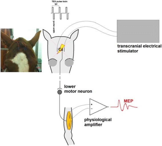
Background: There are indications that transcranial electrical stimulation (TES) assesses the motor function of the spinal cord in horses in a more sensitive and reproducible fashion than transcranial magnetic stimulation (TMS). However, no normative data of TES evoked motor potentials (MEP) is available.
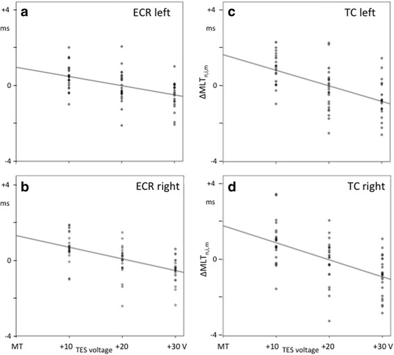
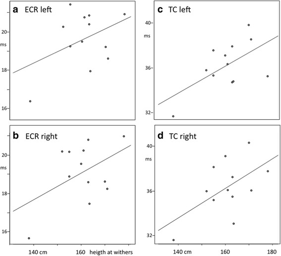
Results: In this prospective study normative data of TES induced MEP wave characteristics (motor latency times (MLT); amplitude and waveform) was obtained from the extensor carpi radialis (ECR) and tibial cranialis (TC) muscles in a group of healthy horses to create a reference frame for functional diagnostic purposes. For the 12 horses involved in the study 95% confidence intervals for MLTs were 16.1-22.6 ms and 31.9-41.1 ms for ECR and TC muscles respectively. Intra-individual coefficients of variation (CV) and mean of MLTs were: ECR: 2.2-8,2% and 4.5% and TC: 1.4-6.3% and 3.5% respectively. Inter-individual CVs for MLTs were higher, though below 10% on all occasions. The mean ± sd of MEP amplitudes was respectively 3.61 ± 2.55 mV (ECR muscle left) and 4.53 ± 3.1 mV (right) and 2.66 ± 2.22 mV (TC muscle left) and 2.55 ± 1.85 mV (right). MLTs showed no significant left versus right differences. All MLTs showed significant (p < 0.05) voltage dependent decreases with slope coefficients of linear regression for ECR: - 0.049; - 0.061 ms/V and TC: - 0.082; - 0.089 ms/V (left; right). There was a positive correlation found between height at withers and MLTs in all 4 muscle groups. Finally, reliable assessment of MEP characteristics was for all muscle groups restricted to a transcranial time window of approximately 15-19 ms.
Conclusions: TES is a novel and sensitive technique to assess spinal motor function in horses. It is easy applicable and highly reproducible. This study provides normative data in healthy horses on TES induced MEPs in the extensor carpi radialis and tibialis cranialis muscles bilaterally. No significant differences between MLTs of the left and right side could be demonstrated. A significant effect of stimulation voltage on MLTs was found. No significant effect of height at the withers could be found based upon the results of the current study. A study in which both TMS and TES are applied on the same group of horses is needed.
Keywords: Horses; MEP; Neurology; Spinal cord function; TES; TMS; Transcranial electric stimulation.
Conflict of interest statement
Ethics approval
The study was approved by the Animal Ethics Committee of University of Groningen, The Netherlands under the ethical committee reference DEC6440A, including signed Informed consent from the horse owners.
Consent for publication
Not applicable for this study.
Competing interests
The authors declare that they have no competing interests.
Publisher’s Note
Springer Nature remains neutral with regard to jurisdictional claims in published maps and institutional affiliations.
Figures
Similar articles
-
Trapezius Motor Evoked Potentials From Transcranial Electrical Stimulation and Transcranial Magnetic Stimulation: Reference Data, Characteristic Differences and Intradural Motor Velocities in Horses.Front Neurosci. 2022 Apr 27;16:851463. doi: 10.3389/fnins.2022.851463. eCollection 2022. Front Neurosci. 2022. PMID: 35573305 Free PMC article.
-
Comparison of Muscle MEPs From Transcranial Magnetic and Electrical Stimulation and Appearance of Reflexes in Horses.Front Neurosci. 2020 Sep 25;14:570372. doi: 10.3389/fnins.2020.570372. eCollection 2020. Front Neurosci. 2020. PMID: 33122992 Free PMC article.
-
Transcranial magnetic stimulation: normal values of magnetic motor evoked potentials in 84 normal horses and influence of height, weight, age and sex.Equine Vet J. 2004 Jan;36(1):51-7. doi: 10.2746/0425164044864660. Equine Vet J. 2004. PMID: 14756372
-
State-of-the-Art Diagnostic Methods to Diagnose Equine Spinal Disorders, With Special Reference to Transcranial Magnetic Stimulation and Transcranial Electrical Stimulation.J Equine Vet Sci. 2019 Oct;81:102790. doi: 10.1016/j.jevs.2019.102790. Epub 2019 Sep 3. J Equine Vet Sci. 2019. PMID: 31668311 Review.
-
Transcranial Magnetic Stimulation and Transcranial Electrical Stimulation in Horses.Vet Clin North Am Equine Pract. 2022 Aug;38(2):189-211. doi: 10.1016/j.cveq.2022.04.002. Epub 2022 Jul 7. Vet Clin North Am Equine Pract. 2022. PMID: 35811197 Review.
Cited by
-
Evaluation of the diagnostic value of transcranial electrical stimulation (TES) to assess neuronal functional integrity in horses.Front Neurosci. 2024 Apr 11;18:1342803. doi: 10.3389/fnins.2024.1342803. eCollection 2024. Front Neurosci. 2024. PMID: 38665290 Free PMC article.
-
Trapezius Motor Evoked Potentials From Transcranial Electrical Stimulation and Transcranial Magnetic Stimulation: Reference Data, Characteristic Differences and Intradural Motor Velocities in Horses.Front Neurosci. 2022 Apr 27;16:851463. doi: 10.3389/fnins.2022.851463. eCollection 2022. Front Neurosci. 2022. PMID: 35573305 Free PMC article.
-
Comparison of Muscle MEPs From Transcranial Magnetic and Electrical Stimulation and Appearance of Reflexes in Horses.Front Neurosci. 2020 Sep 25;14:570372. doi: 10.3389/fnins.2020.570372. eCollection 2020. Front Neurosci. 2020. PMID: 33122992 Free PMC article.
-
Extramuscular Recording of Spontaneous EMG Activity and Transcranial Electrical Elicited Motor Potentials in Horses: Characteristics of Different Subcutaneous and Surface Electrode Types and Practical Guidelines.Front Neurosci. 2020 Jul 17;14:652. doi: 10.3389/fnins.2020.00652. eCollection 2020. Front Neurosci. 2020. PMID: 32765207 Free PMC article.
References
MeSH terms
LinkOut - more resources
Full Text Sources
Other Literature Sources
