. 2018 Apr 3;9(1):81.
doi: 10.1186/s13287-018-0819-z.

Human chorionic plate-derived mesenchymal stem cells transplantation restores ovarian function in a chemotherapy-induced mouse model of premature ovarian failure

Affiliations

Human chorionic plate-derived mesenchymal stem cells transplantation restores ovarian function in a chemotherapy-induced mouse model of premature ovarian failure

Jun Li et al. Stem Cell Res Ther. .

Abstract

Background: Previous studies have reported that transplantation of mesenchymal stem cells (MSCs) from many human tissues could ameliorate ovarian dysfunction. However, no study has revealed the therapeutic efficiency of MSCs derived from the chorionic plate (CP-MSCs) for premature ovarian failure (POF).

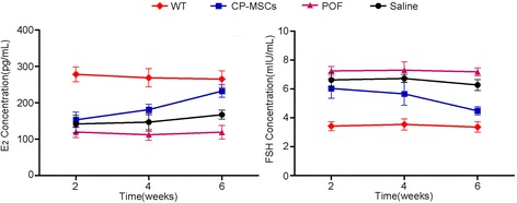
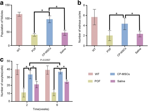
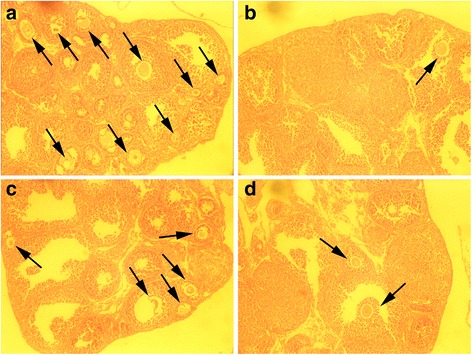
Methods: We investigated the restorative effects of CP-MSCs on cyclophosphamide (CTX)-induced POF. The POF mouse models were established via intraperitoneal injection of 50 mg/kg CTX into female mice for 15 consecutive days. After that, CP-MSCs were intravenously transplanted into the mice once a week for 4 weeks. The serum estradiol (E2) and follicle-stimulating hormone (FSH) levels in the mouse models were detected using enzyme-linked immunosorbent assay (ELISA) before and after treatment. Ovarian function was evaluated through counting the follicles, estrous cycles, and oocytes.

Results: CP-MSC transplantation restored the serum hormone level and ovarian function of the mice in the mouse model of POF induced by CTX. The levels of serum E2 and FSH in the POF model group was 232.33 ± 17.16 pg/mL and 4.48 ± 0.29 mIU/mL, respectively, after 6 weeks of treatment, which were similar to the values in the wild-type (WT) group. The superovulation demonstrated that ovarian function was significantly improved compared with nontreated POF model mice. The CP-MSC transplantation could restore CTX-induced ovarian dysfunction.

Conclusions: Our results offer a potential application for human CP-MSCs in POF treatment.

Keywords: CP-MSCs; Cyclophosphamide; Ovarian function; Premature ovarian failure; Transplantation.


Conflict of interest statement

Ethics approval and consent to participate

Human placental tissue was kindly provided by the Affiliated Hospital of Jiangsu University in accordance with the Declaration of Helsinki. All animal procedures were approved by the ethics committee of Jiangsu University in accordance with the Principe of Laboratory Animal Care (NIH Publication No. 85-23, revised 1985).

Consent for publication

All authors agree to the publication of this article.

Competing interests

The authors declare that they have no competing interests.

Publisher’s Note

Springer Nature remains neutral with regard to jurisdictional claims in published maps and institutional affiliations.

Figures

Fig. 1
Fig. 1
Preparation and karyotyping of CP-MSCs. CP-MSCs were isolated using the explants method (a) and expanded to the fifth passage (bf, passage one to passage five). Scale bars = 100 μm. g Karyotyping was carried out on passage 5 CP-MSCs, and no significant chromosome variation was observed
Fig. 2
Fig. 2
Flow cytometry and secretion of growth factors. a Passage 3 CP-MSCs were analyzed by flow cytometry to show the expression of surface markers CD11b, CD19, CD34, CD45, HLA-DR, CD29, CD44, CD73, CD90, and CD105. CP-MSCs were positive for CD29, CD44, CD73, CD90, and CD105, and negative for CD11b, CD19, CD34, CD45, and HLA-DR. b,c Levels of the growth factors hepatocyte growth factor (HGF), platelet derived growth factor (PDGF), insulin-like growth factor (IGF)-1, vascular endothelial growth factor (VEGF), transforming growth factor (TGF), fibroblast growth factor (FGF), and epidermal growth factor (EGF) were determined. The CP-MSCs secreted high levels of IGF-1, PDGF, and HGF, medium levels of TGF, low levels of FGF and EGF, and scarcely secreted VEGF
Fig. 3
Fig. 3
Differentiation of CP-MSCs. a Blank control; scale bar = 100 μm. CP-MSCs at passage 3 were induced to adipogenesis (b), chondrogenesis (c), and osteogenesis (d); scale bars = 50 μm. CP-MSCs possessed the potential for differentiation to adipocytes containing scattered lipid droplets which were stained red with Oil Red O (b), to chondrocytes which were stained blue with Alcian Blue (c), and to osteocytes with formation of calcium nodules which were stained red with Alizarin Red (d)
Fig. 4
Fig. 4
Improvement in hormone levels after placental CP-MSC transplantation. A significant improvement in the hormone level was observed in premature ovarian failure (POF) after the administration of placental chorionic plate-derived mesenchymal stem cells (CP-MSCs) (P < 0.05, CP-MSC treatment group versus the POF model group). The administration of saline to the POF mouse model improved the hormone level slightly; however, this change was not significant (P > 0.05, saline treatment group versus POF model group). The levels of estradiol (E2) and follicle-stimulating hormone (FSH) in the serum was assayed at 2, 4, and 6 weeks post-transplantation of CP-MSCs. The hormone concentration of wild-type (WT) and POF model groups were maintained at a stable level (P < 0.05, POF model group versus WT group). WT: wild-type mouse without any treatment as a blank control group; CP-MSCs: POF mouse model treated with CP-MSCs as an experimental group; POF: POF mouse model without any treatment as a negative control group; Saline: POF mouse model treated with saline as a vehicle group
Fig. 5
Fig. 5
The function of the ovary in the POF mouse model improved after CP-MSC transplantation. a The population of follicles in the premature ovarian failure (POF) model mouse increased significantly after treatment with chorionic plate-derived mesenchymal stem cells (CP-MSCs) compared with the POF mouse model treated with saline. b The estrous cycles of the mice were counted post-treatment. c The oocytes of mice in each group were measured via superovulation to validate the ovarian function. (*P < 0.05) WT: wild-type mouse without any treatment as a blank control group; CP-MSCs: POF mouse model treated with CP-MSCs as an experimental group; POF: POF mouse model without any treatment as a negative control group; Saline: POF mouse model treated with saline as a vehicle group
Fig. 6
Fig. 6
Histological analysis of mouse ovaries. Six weeks after treatment, mice ovaries were collected and fixed, followed by dehydration, paraffin embedding, and serial sectioning. The sections were stained with hematoxylin and eosin. a WT group, b POF mouse model without any treatment, c POF mouse model treated with CP-MSCs, and d POF mouse model treated with saline. Original magnification: 100×. Black arrows indicate the follicles containing a clear oocyte

References

    1. Torrealday S, Pal L. Premature menopause. Endocrinol Metab Clin North Am. 2015;44:543–557. doi: 10.1016/j.ecl.2015.05.004. - DOI - PubMed
    1. Bilgin EM, Kovanci E. Genetics of premature ovarian failure. Curr Opin Obstet Gynecol. 2015;27:167–174. doi: 10.1097/GCO.0000000000000177. - DOI - PubMed
    1. Laven JSE. Primary ovarian insufficiency. Semin Reprod Med. 2016;34:230–234. doi: 10.1055/s-0036-1585402. - DOI - PubMed
    1. Kovanci E, Schutt AK. Premature ovarian failure: clinical presentation and treatment. Obstet Gynecol Clin North Am. 2015;42:153–161. doi: 10.1016/j.ogc.2014.10.004. - DOI - PubMed
    1. Lee H-J, Selesniemi K, Niikura Y, Niikura T, Klein R, Dombkowski DM, Tilly JL. Bone marrow transplantation generates immature oocytes and rescues long-term fertility in a preclinical mouse model of chemotherapy-induced premature ovarian failure. J Clin Oncol. 2007;25:3198–3204. doi: 10.1200/JCO.2006.10.3028. - DOI - PubMed

Publication types

Substances