. 2018 Apr 3;9(1):1314.
doi: 10.1038/s41467-018-03562-9.

Rescue of conformational dynamics in enzyme catalysis by directed evolution

Affiliations

Rescue of conformational dynamics in enzyme catalysis by directed evolution

Renee Otten et al. Nat Commun. .

Abstract

Rational design and directed evolution have proved to be successful approaches to increase catalytic efficiencies of both natural and artificial enzymes. Protein dynamics is recognized as important, but due to the inherent flexibility of biological macromolecules it is often difficult to distinguish which conformational changes are directly related to function. Here, we use directed evolution on an impaired mutant of the proline isomerase CypA and identify two second-shell mutations that partially restore its catalytic activity. We show both kinetically, using NMR spectroscopy, and structurally, by room-temperature X-ray crystallography, how local perturbations propagate through a large allosteric network to facilitate conformational dynamics. The increased catalysis selected for in the evolutionary screen is correlated with an accelerated interconversion between the two catalytically essential conformational sub-states, which are both captured in the high-resolution X-ray ensembles. Our data provide a glimpse of an evolutionary trajectory and show how subtle changes can fine-tune enzyme function.


Conflict of interest statement

The authors declare no competing interests.

Figures

Fig. 1
Fig. 1
Directed evolution selects rescue mutations for catalysis. a Scheme of the assay used in directed evolution to identify CypA mutations with increased catalytic activity: CypA activity for folding of AvrRpt2 protease measured by AvrRpt2-mediated cleavage of the peptide Abz-IEAPAFGGWy-NH2 (y = 3-nitro-Tyrosine). b Assay of directed evolution performed on cell lysate in 96-well plates to identify rescue mutations for S99T with increased CypA activity. Kinetics of peptide cleavage is shown for wild-type (yellow), S99T (green), and S99T/C115S (blue) CypA. c, d The severely catalytically compromised S99T mutant (green) is rescued by second-shell mutations (C115S, blue and I97V, red). c Sites of mutations are plotted onto the crystal structure (1RMH) of CypA bound to Suc-AAPF-pNA (gray sticks) and the active-site residues Arg55 and Phe113 are shown in black stick representation. The overlay of NMR spectra shows that the overall structure of all CypA forms is very similar, with perturbations observed for residues close to the mutation site or in the dynamic network (Supplementary Fig. 1). d kcat/KM values for wild-type, S99T, S99T/C115S, and S99T/C115S/I97V CypA measured by protease coupled hydrolysis of Suc-AFPF-pNA and Suc-AAPF-pNA peptides (see Table 1). Error bars indicate the standard deviation obtained from triplicate measurements on at least three different enzyme concentrations. e KD values for the three mutant forms of CypA for Suc-AFPF-pNA measured by NMR chemical shift analysis from peptide titrations (see also Supplementary Fig. 2 and Table 1). Error bars denote the standard errors in the fitted parameters obtained from the global fit
Fig. 2
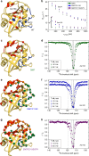
Fig. 2
Rescue mutants alter the conformational dynamics of CypA as measured by NMR. a Dynamics on WT CypA (shown here) and S99T (c) identified a slower (group-I, red) and faster dynamical process (group-II, blue). Residues Gly74 (blue) and Asn102 (red) are indicated with a sphere. b For the three mutants, 15N-CPMG dispersion profiles for a representative residue in fast exchange (Gly74, group-II) and slow exchange (Asn102, group-I). The fast-exchange process is virtually unaltered by the mutations, whereas Rex increases consecutively from single via double to triple-mutant (see also Supplementary Data 3). c, e, g Quantitative analysis of fast and slow protein dynamics of CypA rescue mutants by CPMG relaxation and CEST experiments are plotted onto the structure. Blue and red color coding of the cartoon representation denotes fast and slow dynamics, respectively, as determined from the temperature-dependence and shape of CPMG relaxation dispersion profiles (Supplementary Data 1 and 2). Spheres represent residues in slow exchange with quantifiable CEST profiles. c CEST data for all 15 residues (red spheres) in S99T can be globally fit to a two-site exchange process (Supplementary Fig. 3, Supplementary Data 4 and Table 2). e, g Residues with CEST profiles in S99T/C115S (e) and S99T/C115S/I97V (g) are well-described by two distinct slow processes (red and green, respectively), whereas residues shown in orange sense both processes and require a three-site exchange model (Supplementary Figs. 4 and 5, Supplementary Data 5 and 6 and Table 2). d, f, h Representative 15N-CEST profiles of residue Asn102, measured at the indicated field strengths are shown for single (d), double (f), and triple (h) mutants of CypA. The chemical shifts for the major (solid line) and minor states (– – – and – · – lines) are indicated. Uncertainties in R2,eff (b) are determined from the rmsd in the intensities of duplicate points (n = 4) according to the definition of pooled relative standard deviation; uncertainties in I/I0 for CEST data (d, f, h) are determined from the rmsd in the baseline of the profile where no intensity dips are present (typically, n > 50)
Fig. 3
Fig. 3
Protein dynamics during turnover and catalytic efficiency correlate. a Correlation between kcat/KM and kmaj→min measured by CEST across all rescue mutations for apo protein fitted using orthogonal distance regression through the origin (AFPF: adj. R2 = 0.999, χ2red = 1.1; AAPF: adj. R2 = 0.995, χ2red = 3.7). b Correlation of the chemical shift differences between major and minor conformations for the two processes observed in S99T/C115S and S99T/C115S/I97V. Residues within 5 Å of mutation site (I97V) are shown in black and assignments are given if the variation in Δδ is >1.5 ppm. c Quantitative analysis of fast and slow protein dynamics of CypA S99T during catalysis of Suc-AFPF-pNA peptide (gray sticks) measured by NMR. CPMG relaxation dispersion experiments revealed, similarly as in apo S99T, fast motion mainly in the flexible loop (blue) and a slow process (red) consistent with the CEST data (spheres). d Representative CPMG profiles for a residue in fast (Gly74) and slow (Glu120) exchange and e CEST profile for Glu120 during catalysis (see Supplementary Data 7 and 8 for all profiles). Error bars in a and b denote the (propagated) standard errors in the fitted parameters. The uncertainties in CPMG (d) and CEST (e) data are determined as described in Fig. 2
Fig. 4
Fig. 4
Structural basis of the increased protein dynamics from room-temperature X-ray crystallography on rescue mutants. a X-ray structure of CypA (2CPL) with the active-site residues Arg55, Ser99, and Phe113 shown in black stick representation. The boxed area indicates the extended dynamic network shown in more detail in b and c. b Major and minor side chain conformations are shown for S99T/C115S/I97V (5WC7, 1.43 Å, see Supplementary Table 1) in purple and pink, respectively. The populations are flipped relative to wild-type CypA (3K0N, 1.4 Å) (c), where major/minor states are shown in red and orange, respectively. The Phe113-in state corresponds to the major WT state and the minor S99T/C115S/I97V state. Correspondingly, the Phe113-out state corresponds to the minor WT state and the major S99T/C115S/I97V state. d, e, f Less steric hindrance due to the reduced size of side chains in rescue mutants facilitates the interconversion between major and minor conformations. Coupling between the conformation of Thr99 and Phe113 (d) and Val97 and Thr99 (e) are necessary to relieve clashes. The C115S (f, purple) mutation allows for a transition between Thr99 conformations (gray indicates morph between the major in minor state labeled Thr99*) without clash, in contrast to the bulkier Cys residue (green). g CONTACT analysis of alternative conformations of S99T/C115S/I97V identifies a network extending across group-I residues (pink surface representation) consistent with the NMR results

References

    1. Kay LE. New views of functionally dynamic proteins by solution NMR spectroscopy. J. Mol. Biol. 2016;428:323–331. doi: 10.1016/j.jmb.2015.11.028. - DOI - PubMed
    1. Boehr DD, Dyson HJ, Wright PE. An NMR perspective on enzyme dynamics. Chem. Rev. 2006;106:3055–3079. doi: 10.1021/cr050312q. - DOI - PubMed
    1. Smock RG, Gierasch LM. Sending signals dynamically. Science. 2009;324:198–203. doi: 10.1126/science.1169377. - DOI - PMC - PubMed
    1. Palmer AG., 3rd Enzyme dynamics from NMR spectroscopy. Acc. Chem. Res. 2015;48:457–465. doi: 10.1021/ar500340a. - DOI - PMC - PubMed
    1. Shukla D, Hernandez CX, Weber JK, Pande VS. Markov state models provide insights into dynamic modulation of protein function. Acc. Chem. Res. 2015;48:414–422. doi: 10.1021/ar5002999. - DOI - PMC - PubMed

Publication types