

Protective Role of Perivascular Adipose Tissue in Endothelial Dysfunction and Insulin-Induced Vasodilatation of Hypercholesterolemic LDL Receptor-Deficient Mice
- PMID: 29615924
- PMCID: PMC5868473
- DOI: 10.3389/fphys.2018.00229
Protective Role of Perivascular Adipose Tissue in Endothelial Dysfunction and Insulin-Induced Vasodilatation of Hypercholesterolemic LDL Receptor-Deficient Mice
Abstract
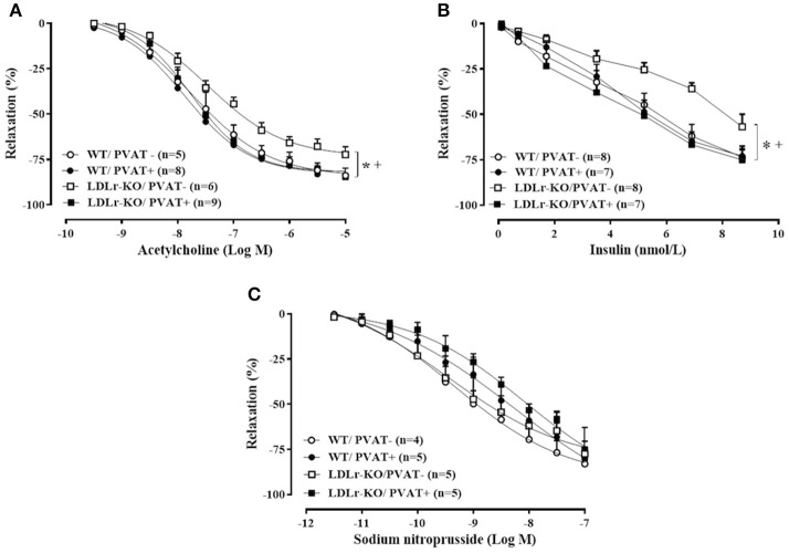
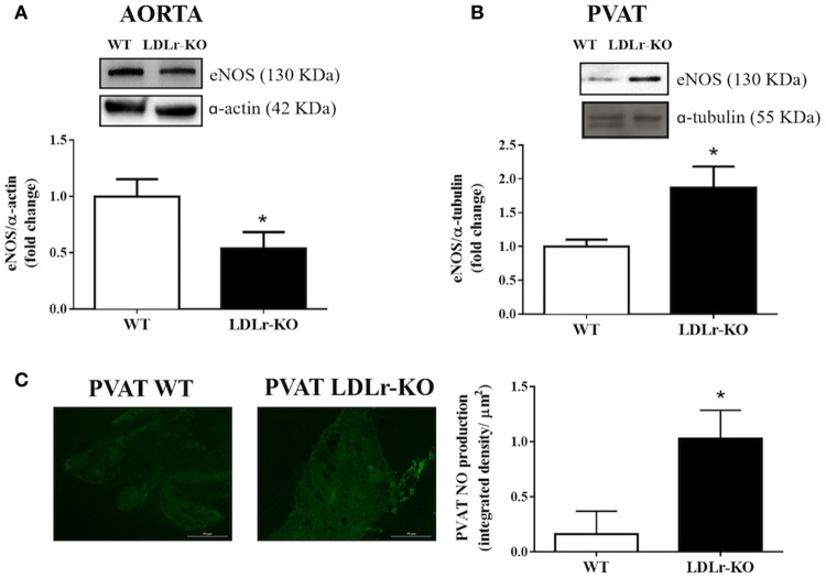
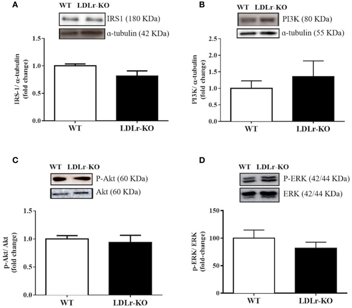
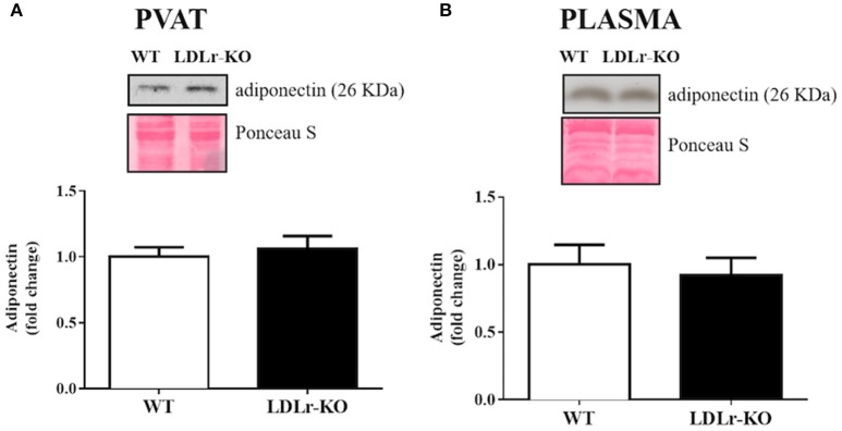
Background: Endothelial dysfunction plays a pivotal role in the initiation of atherosclerosis. Vascular insulin resistance might contribute to a reduction in endothelial nitric oxide (NO) production, leading to impaired endothelium-dependent relaxation in cardiometabolic diseases. Because perivascular adipose tissue (PVAT) controls endothelial function and NO bioavailability, we hypothesized a role for this fat deposit in the vascular complications associated with the initial stages of atherosclerosis. Therefore, we investigated the potential involvement of PVAT in the early endothelial dysfunction in hypercholesterolemic LDL receptor knockout mice (LDLr-KO). Methods: Thoracic aortas with and without PVAT were isolated from 4-month-old C57BL/6J (WT) and LDLr-KO mice. The contribution of PVAT to relaxation responses to acetylcholine, insulin, and sodium nitroprusside was investigated. Western blotting was used to examine endothelial NO synthase (eNOS) and adiponectin expression, as well the insulin signaling pathway in aortic PVAT. Results: PVAT-free aortas of LDLr-KO mice exhibited impaired acetylcholine- and insulin-induced relaxation compared with those of WT mice. Both vasodilatory responses were restored by the presence of PVAT in LDLr-KO mice, associated with enhanced acetylcholine-induced NO levels. PVAT did not change vasodilatory responses to acetylcholine and insulin in WT mice, while vascular relaxation evoked by the NO donor sodium nitroprusside was not modified by either genotype or PVAT. The expression of insulin receptor substrate-1 (IRS-1), phosphatidylinositol 3-kinase (PI3K), AKT, ERK1/2, phosphorylation of AKT (Ser473) and ERK1/2 (Thr202/Tyr204), and adiponectin was similar in the PVAT of WT and LDLr-KO mice, suggesting no changes in PVAT insulin signaling. However, eNOS expression was enhanced in the PVAT of LDLr-KO mice, while eNOS expression was less abundant in PVAT-free aortas. Conclusion: These results suggest that elevated eNOS-derived NO production in aortic PVAT might be a compensatory mechanism for the endothelial dysfunction and impaired vasodilator action of insulin in hypercholesterolemic LDLr-deficient mice. This protective effect may limit the progression of atherosclerosis in genetic hypercholesterolemia in the absence of an atherogenic diet.
Keywords: LDL receptor deficiency; adiponectin; endothelium; hypercholesterolemia; insulin; nitric oxide; perivascular adipose tissue.
Figures
References
-
- Bonfleur M. L., Ribeiro R. A., Balbo S. L., Vanzela E. C., Carneiro E. M., de Oliveira H. C., et al. (2011). Lower expression of PKAα impairs insulin secretion in islets isolated from low-density lipoprotein receptor (LDLR−/−) knockout mice. Metabolism 60, 1158–1164. 10.1016/j.metabol.2010.12.010 - DOI - PubMed
LinkOut - more resources
Full Text Sources
Other Literature Sources
Molecular Biology Databases
Research Materials
Miscellaneous