Clinical Trial
. 2018 Mar 19:9:472.
doi: 10.3389/fimmu.2018.00472. eCollection 2018.

Hyper-Expression of PD-1 Is Associated with the Levels of Exhausted and Dysfunctional Phenotypes of Circulating CD161++TCR iVα7.2+ Mucosal-Associated Invariant T Cells in Chronic Hepatitis B Virus Infection

Affiliations
Clinical Trial

Hyper-Expression of PD-1 Is Associated with the Levels of Exhausted and Dysfunctional Phenotypes of Circulating CD161++TCR iVα7.2+ Mucosal-Associated Invariant T Cells in Chronic Hepatitis B Virus Infection

Yean K Yong et al. Front Immunol. .

Abstract

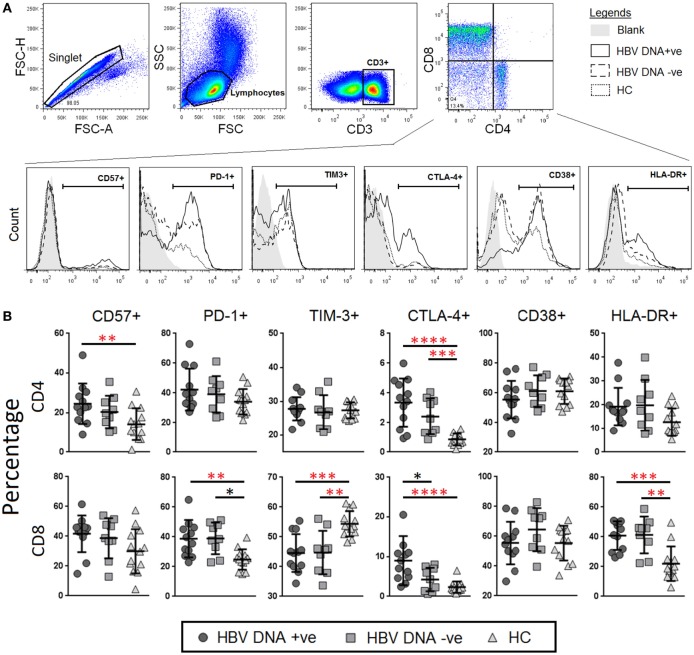
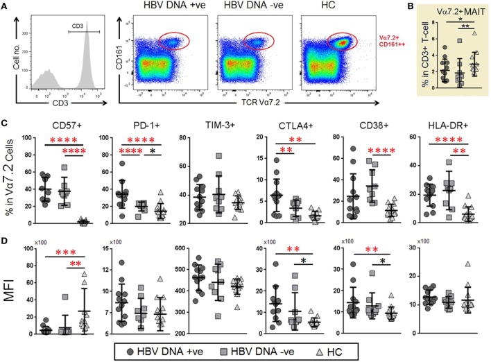
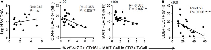
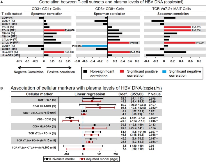
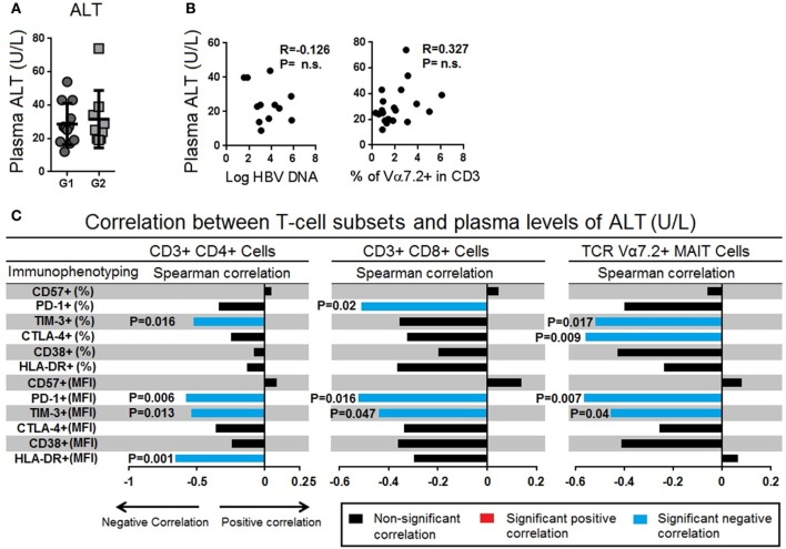
Mucosal-associated invariant T (MAIT) cells, defined as CD161++TCR iVα7.2+ T cells, play an important role in the innate defense against bacterial infections, and their functionality is impaired in chronic viral infections. Here, we investigated the frequency and functional role of MAIT cells in chronic hepatitis B virus (HBV) infection. The peripheral CD3+CD161++TCR iVα7.2+ MAIT cells in chronic HBV-infected patients and healthy controls were phenotypically characterized based on CD57, PD-1, TIM-3, and CTLA-4, as well as HLA-DR and CD38 expression. The frequency of MAIT cells was significantly decreased among chronic HBV-infected individuals as compared to controls. Expression of CD57, PD-1, CTLA-4, as well as HLA-DR and CD38 on MAIT cells was significantly elevated in chronic HBV-infected individuals relative to controls. The percentage of T cell receptor (TCR) iVα7.2+ CD161+ MAIT cells did not correlate with HBV viral load but inversely with HLA-DR on CD4+ T cells and MAIT cells and with CD57 on CD8+ T cells suggesting that decrease of MAIT cells may not be attributed to direct infection by HBV but driven by HBV-induced chronic immune activation. The percentage and expression levels of PD-1 as well as CTLA-4 on MAIT cells inversely correlated with plasma HBV-DNA levels, which may suggest either a role for MAIT cells in the control of HBV infection or the effect of HBV replication in the liver on MAIT cell phenotype. We report that decrease of TCR iVα7.2+ MAIT cells in the peripheral blood and their functions were seemingly impaired in chronic HBV-infected patients likely because of the increased expression of PD-1.

Keywords: CTLA-4; HBV infection; HLA-DR; PD-1; immune exhaustion; immunosenescence; mucosal-associated invariant T cells.


Figures

Figure 1
Figure 1
Frequencies and levels of immune senescence, exhaustion, and activation markers expressed on CD4+ and CD8+ T-cells between chronic hepatitis B virus (HBV)-infected patients with (circle) and without (square) HBV-DNAemia and healthy control (HC) (triangle). (A) The gating strategy to identify (B) expression levels of CD57, PD-1, TIM-3, CTLA-4 CD38, and HLA-DR on CD4+ and CD8+ T-cells. Levels of surface markers were compared across the three patient groups and post hoc Mann–Whitney U tests were then performed for those biomarkers with a Kruskal–Wallis test P value of <0.05 (*P < 0.05, **<0.01, ***<0.001, and ****<0.0001). P-values remained significant after Benjamini–Hochberg correction of multiple comparisons (marked in red*).
Figure 2
Figure 2
Frequencies and levels of immune senescence, exhaustion, and activation markers expressed on mucosal-associated invariant T (MAIT) cells between chronic hepatitis B virus (HBV)-infected patients with (G1, circle) and without (G2, square) HBV-DNAemia and healthy control (HC) (G3, triangle). (A) The gating strategy to identify CD3+TCR Vα7.2+ MAIT cells and Vα7.2+CD161 T cells. (B) Comparison of T cell receptor (TCR) Vα7.2+ cells frequencies across the three groups comparison of percentage. (C) Expression levels [mean fluorescence intensity (MFI)] (D) of CD57, PD-1, TIM-3, CTLA-4 CD38 and HLA-DR on TCR Vα7.2+ MAIT cells between the three patient groups. Levels of surface markers were compared across the three patient groups and post hoc Mann–Whitney U tests were then performed for those biomarkers with a Kruskal–Wallis test P value of <0.05 (*P < 0.05, **<0.01, ***<0.001, and ****<0.0001). P-values remained significant after Benjamini–Hochberg correction of multiple comparisons (marked in red*).
Figure 3
Figure 3
Spearman correlation between frequencies of T cell receptor Vα7.2+ CD161+ mucosal-associated invariant T (MAIT) with (A) markers of immune activation and (B) markers of immune senescence.
Figure 4
Figure 4
(A) Spearman correlations between surface markers in CD4+ (left panel), CD8+ (middle panel), and mucosal-associated invariant T (MAIT) cells (right panel) with level of plasma hepatitis B virus (HBV)-DNA. The bar represents the strength of association (r values) where red bar represents significant positive correlation, blue bar represent significant negative association and black bar represents P value > 0.05 (non-significant association) (<0.05, **<0.01, ***<0.001, and ****<0.0001). (B) Association of all surface markers that showed significant correlation with plasma HBV-DNA levels were assessed in simple logistic regression model and adjusted for age. Coefficient values below or above threshold levels were displayed in a forest plot; median and 95% CI were calculated. CI, confidence interval (*P < 0.05).
Figure 5
Figure 5
(A) Comparison of serum alanine transaminase (ALT) levels between chronic hepatitis B virus (HBV)-infected patients with HBV-DNAemia (circle) and without HBV-DNAemia (square). (B) Spearman correlation between serum ALT levels and frequencies of T cell receptor (TCR) Vα7.2+ CD161+ mucosal-associated invariant T (MAIT) cells. (C) Spearman correlation between frequencies and expression levels of CD57, PD-1, TIM-3, CTLA-4 CD38, and HLA-DR with serum level of ALT. The bar represents the strength of association (r values) where red bar represents significant positive correlation, blue bar represent significant negative association and black bar represents P value > 0.05 (non-significant association) (<0.05, **<0.01, ***<0.001, and ****<0.0001).
Figure 6
Figure 6
(A) Representative density dot plots for perforin, granzyme-B, IFN-γ, and TNF-α (without stimulation) in T cell receptor (TCR) Vα7.2+ mucosal-associated invariant T (MAIT) cells in chronic hepatitis B virus (HBV)-infected patients with HBV-DNAemia (G1) and without HBV-DNAemia (G2) and healthy control (HC) (G3). (B) Comparisons of percentages of perforin, granzyme-B, IFN-γ, and TNF-α in TCR Vα7.2+ across the three groups. (C) Comparisons of expression levels [mean fluorescence intensity (MFI)] of perforin, granzyme-B, IFN-γ, and TNF-α in TCR Vα7.2+ before and after PMA stimulation. (D) Expression (measured as fold change of MFI) of perforin, granzyme-B, IFN-γ, and TNF-α in TCR Vα7.2+ after PMA stimulation. Levels of cytokine expression were compared across the three patient groups and post hoc Mann–Whitney U tests were subsequently performed for those biomarkers using a Kruskal–Wallis test (level of significance P < 0.05) (*P < 0.05, **<0.01, ***<0.001, and ****<0.0001). P-values remained significant after Benjamini–Hochberg correction of multiple comparisons (marked in red*).

References

    1. Lozano R, Naghavi M, Foreman K, Lim S, Shibuya K, Aboyans V, et al. Global and regional mortality from 235 causes of death for 20 age groups in 1990 and 2010: a systematic analysis for the Global Burden of Disease Study 2010. Lancet (2012) 380(9859):2095–128.10.1016/S0140-6736(12)61728-0 - DOI - PMC - PubMed
    1. Bertoletti A, Gehring AJ. Immune therapeutic strategies in chronic hepatitis B virus infection: virus or inflammation control? PLoS Pathog (2013) 9(12):e1003784.10.1371/journal.ppat.1003784 - DOI - PMC - PubMed
    1. Pol S, Nalpas B, Driss F, Michel ML, Tiollais P, Denis J, et al. Efficacy and limitations of a specific immunotherapy in chronic hepatitis B. J Hepatol (2001) 34(6):917–21.10.1016/S0168-8278(01)00028-9 - DOI - PubMed
    1. Vandepapeliere P, Lau GK, Leroux-Roels G, Horsmans Y, Gane E, Tawandee T, et al. Therapeutic vaccination of chronic hepatitis B patients with virus suppression by antiviral therapy: a randomized, controlled study of co-administration of HBsAg/AS02 candidate vaccine and lamivudine. Vaccine (2007) 25(51):8585–97.10.1016/j.vaccine.2007.09.072 - DOI - PubMed
    1. Perrillo R. Benefits and risks of interferon therapy for hepatitis B. Hepatology (2009) 49(5 Suppl):S103–11.10.1002/hep.22956 - DOI - PubMed

Publication types

MeSH terms

Substances