. 2018 Mar 20:9:364.
doi: 10.3389/fpls.2018.00364. eCollection 2018.

The Rice Rolled Fine Striped (RFS) CHD3/Mi-2 Chromatin Remodeling Factor Epigenetically Regulates Genes Involved in Oxidative Stress Responses During Leaf Development

Affiliations

The Rice Rolled Fine Striped (RFS) CHD3/Mi-2 Chromatin Remodeling Factor Epigenetically Regulates Genes Involved in Oxidative Stress Responses During Leaf Development

Sung-Hwan Cho et al. Front Plant Sci. .

Abstract

In rice (Oryza sativa), moderate leaf rolling increases photosynthetic competence and raises grain yield; therefore, this important agronomic trait has attracted much attention from plant biologists and breeders. However, the relevant molecular mechanism remains unclear. Here, we isolated and characterized Rolled Fine Striped (RFS), a key gene affecting rice leaf rolling, chloroplast development, and reactive oxygen species (ROS) scavenging. The rfs-1 gamma-ray allele and the rfs-2 T-DNA insertion allele of RFS failed to complement each other and their mutants had similar phenotypes, producing extremely incurved leaves due to defective development of vascular cells on the adaxial side. Map-based cloning showed that the rfs-1 mutant harbors a 9-bp deletion in a gene encoding a predicted CHD3/Mi-2 chromatin remodeling factor belonging to the SNF2-ATP-dependent chromatin remodeling family. RFS was expressed in various tissues and accumulated mainly in the vascular cells throughout leaf development. Furthermore, RFS deficiency resulted in a cell death phenotype that was caused by ROS accumulation in developing leaves. We found that expression of five ROS-scavenging genes [encoding catalase C, ascorbate peroxidase 8, a putative copper/zinc superoxide dismutase (SOD), a putative SOD, and peroxiredoxin IIE2] decreased in rfs-2 mutants. Western-blot and chromatin immunoprecipitation (ChIP) assays demonstrated that rfs-2 mutants have reduced H3K4me3 levels in ROS-related genes. Loss-of-function in RFS also led to multiple developmental defects, affecting pollen development, grain filling, and root development. Our results suggest that RFS is required for many aspects of plant development and its function is closely associated with epigenetic regulation of genes that modulate ROS homeostasis.

Keywords: Rolled Fine Striped; chloroplast biogenesis; chromatin remodeling factor; leaf variegation; narrow leaf; reactive oxygen species; rice (Oryza sativa).


Figures
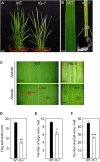
FIGURE 1
FIGURE 1
The rfs-1 mutant has irregular white stripes on the leaf blades. (A) Phenotypes of 2-month-old WT and rfs-1 plants. (B) Rolled-leaf phenotype in the rfs-1 mutant. (C) Adaxial and abaxial sides of WT and rfs-1 leaves. Midrib vein (mv); large vein (lv); small vein (sv). (D) Flag leaf width of WT and rfs-1. (E) Number of large veins in WT and rfs-1 flag leaves. (F) Number of small veins in WT and rfs-1 flag leaves. Scale bars: 5 cm (A); 5 mm (B); 2 mm (C). Error bars indicate ± SD (n = 10). Asterisks indicate statistically significant differences compared to WT, as determined by Student’s t-test (P < 0.05, ∗∗P < 0.01, ∗∗∗P < 0.005).
FIGURE 2
FIGURE 2
The rfs-1 mutants show defective vascular development in leaves. (A,C) Scanning electron micrograph of the WT and rfs-1 adaxial epidermis. bulliform cell (bc); hair (h); trichome (tc). (B,D) Transverse section of WT and rfs-1 large vein. sc (ad), adaxial sclerenchyma; vbs, vascular bundle sheath; m, mesophyll cell; x, xylem; p, phloem; sc (ab), abaxial sclerenchyma. (E,G) Transverse section through WT and rfs-1 small veins. Bulliform cell (bc). (F,H) Enlarged transverse section of WT and rfs-1 small veins. (I,K) Scanning electron micrograph of the WT and rfs-1 abaxial epidermis. large papilla (lp); trichome (tc). (J,L) Enlarged transverse section of WT and rfs-1 bulliform cells. Scale bars: 100 μm (A,C,E,G,I,K); 50 μm (B,D,F,H,J,L).
FIGURE 3
FIGURE 3
The rfs-1 mutant accumulates more ROS in the leaves. (A,B) DAB staining for hydrogen peroxide (H2O2) (dark brown, A) and NBT staining for superoxide (O2-) (blue, B) in the leaf blades of 2-month-old WT and rfs-1. Before, leaf blade prior to staining; DAB, cleared leaf after DAB staining; NBT, cleared leaf after NBT staining. (C) Visualization of singlet oxygen (1O2) detected with the SOSG fluorescent probe on the leaves of 2-month-old WT and rfs-1. The fluorescence of SOSG is shown in green; the red color corresponds to chlorophyll (Chl) auto-fluorescence. Scale bars: 10 μm (B). DAB, 3,3′-diaminobenzidine; NBT, nitroblue tetrazolium; SOSG, singlet oxygen sensor green.
FIGURE 4
FIGURE 4
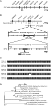
Map-based cloning of RFS and phylogenetic analysis of the RFS proteins in other plants. (A) Genetic mapping of the RFS gene using SSR and STS markers. (B) Physical mapping of the RFS gene. The genomic region contains five expressed genes (gray and black boxes). The genomic structure of RFS, comprising 11 exons and 10 introns, is indicated. A deletion mutation (SFQ 811–813) was found in exon 6 of RFS in the rfs-1 mutant. (C) Nucleotide mutations result in deletion of the amino acids SFQ (asterisks) in the SNF2 family N-terminal domain (gray line). (D) The phylogenetic tree was constructed by the neighbor-joining method with MEGA (ver 5.1) program. Branch numbers represent percentage of bootstrap values in 1000 sampling replicates and the scale indicates branch length.
FIGURE 5
FIGURE 5
Expression profiles of RFS. (A) RT-qPCR analysis of RFS expression in various tissues. Total RNA was isolated from the leaf blade (LB), leaf sheath (LS), shoot base (SB), root (R), and flower (F) of WT. Relative expression levels of RFS were obtained by normalizing to the transcript levels of OsUBQ5. (B–J) Histochemical GUS analysis of the transgenic plants containing the ProRFS:GUS construct. GUS activity was detected in the mesophyll cells (m) and leaf veins of the leaf blade (B–D), especially in the phloem (p) but not in xylem (x) tissues of vascular bundle, longitudinal veins of a spikelet (E), transverse section of leaf blade and node (F,G), vascular cylinder (vc) of primary root (H), and transverse section of large and small vascular bundle in basal node (I,J). Scale bars: 1 mm (B,E,H); 200 μm (C,D); 500 μm (F,G,I,J).
FIGURE 6
FIGURE 6
Altered expression of ROS-related genes in the WT and rfs-2. By RT-qPCR, the expression levels of 21 genes encoding the ROS scavengers were measured in the leaf tissues of 2-month-old WT and rfs-2 (see Supplementary Figure S2C). The five genes whose expression was significantly decreased in the rfs-2 leaves compared with in WT leaves are shown in red type. UBQ5 was used as an internal control for RT-qPCR. CATA; catalase A, CATB; catalase B, CATC, catalase C; APX1, ascorbate peroxidase 1; APX2, ascorbate peroxidase 2; APX3, ascorbate peroxidase 3; APX4, ascorbate peroxidase 4; APX5, ascorbate peroxidase 5; APX6, ascorbate peroxidase 6; APX7, ascorbate peroxidase 7; APX8, ascorbate peroxidase 8; putative Cu/Zn-SOD, a putative copper/zinc superoxide dismutase (SOD); SodCc1, Cu/Zn-SOD 1; putative Cu-SOD, a putative chaperone for copper SOD; SodA1, putative SOD; SodCc2, Cu/Zn-SOD 2; RbohF, NADPH oxidase RbohF; Prx IIE2, peroxiredoxin IIE2. Bars represent ± SD from three independent experiments. Asterisks indicate statistically significant difference compared to WT as determined by Student’s t-test (P < 0.05, ∗∗P < 0.01, ∗∗∗P < 0.005).
FIGURE 7
FIGURE 7
Accumulation of modified histone proteins in the WT and rfs-2. Enriched histone fractions were isolated from 2-week-old WT and rfs-2 seedlings grown in the growth chamber. Western blots were performed by using antibodies against dimethylated H3K9, trimethylated H3K4, and trimethylated H3K27. The blotted membrane was stained with Coomassie Brilliant Blue reagent to show equal loading of each lane. The levels of each protein are shown relative to those of WT, which is set as 1.
FIGURE 8
FIGURE 8
OsRFS modulates H3K4me3 levels on ROS-related genes. Chromatin immunoprecipitation (ChIP) analysis of H3K4me3 on the selected ROS-related genes in the leaves of 2-month-old WT and rfs-2 (see Supplementary Figure S4). Enrichment of H3K4me3 on the promoter (A) and the 5′ end region (B) of the selected ROS-related genes was measured by ChIP followed by qPCR. CATC, APX8, putative Cu/Zn-SOD, putative SOD, Prx IIE2. OsActin7 (ACT) was used for the normalization of the qPCR analysis. Means and standard deviations were obtained from three biological replicates. Asterisks indicate statistically significant difference compared to WT as determined by Student’s t-test (P < 0.05, ∗∗P < 0.01, ∗∗∗P < 0.005).

References

    1. Aasland R., Stewart F. (1995). The chromo shadow domain, a second chromo domain in heterochromatin-binding protein 1, HP1. Nucleic Acids Res. 23 3168–3173. 10.1093/nar/23.16.3168 - DOI - PMC - PubMed
    1. Agrawal G. K., Rakwal R., Jwa N.-S. (2001). Stress signaling molecules involved in defense and protein phosphatase 2A inhibitors modulate OsCATC expression in rice (Oryza sativa) seedlings. J. Plant Physiol. 158 1349–1355. 10.1078/0176-1617-00607 - DOI
    1. Alvarez-Venegas R. (2005). Methylation patterns of histone H3 Lys4, Lys9 and Lys27 in transcriptionally active and inactive Arabidopsis genes and in atx1 mutants. Nucleic Acids Res. 33 5199–5207. 10.1093/nar/gki830 - DOI - PMC - PubMed
    1. Apel K., Hirt H. (2004). Reactive oxygen species: metabolism, oxidative stress, and signal transduction. Annu. Rev. Plant Biol. 55 373–399. 10.1146/annurev.arplant.55.031903.141701 - DOI - PubMed
    1. Asada K. (1999). The water-water cycle in chloroplasts: scavenging of active oxygens and dissipation of excess photons. Annu. Rev. Plant Physiol. Plant Mol. Biol. 50 601–639. 10.1146/annurev.arplant.50.1.601 - DOI - PubMed

LinkOut - more resources