. 2018 Mar 16:8:84.
doi: 10.3389/fcimb.2018.00084. eCollection 2018.

The ATP-P2X7 Signaling Axis Is an Essential Sentinel for Intracellular Clostridium difficile Pathogen-Induced Inflammasome Activation

Affiliations

The ATP-P2X7 Signaling Axis Is an Essential Sentinel for Intracellular Clostridium difficile Pathogen-Induced Inflammasome Activation

Ya-Hui Liu et al. Front Cell Infect Microbiol. .

Erratum in

Abstract

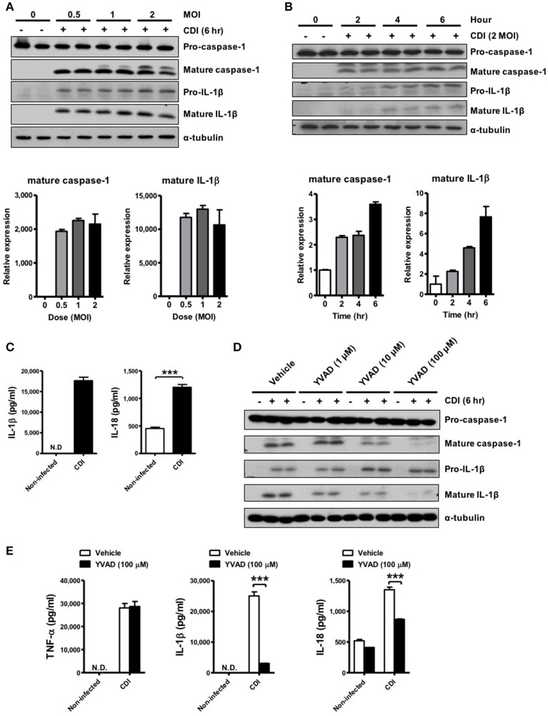
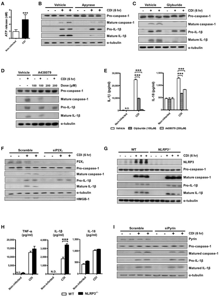
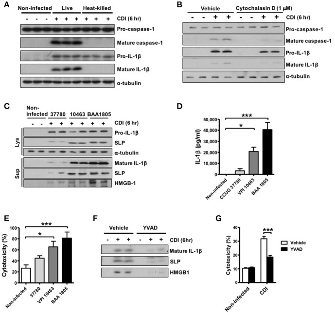
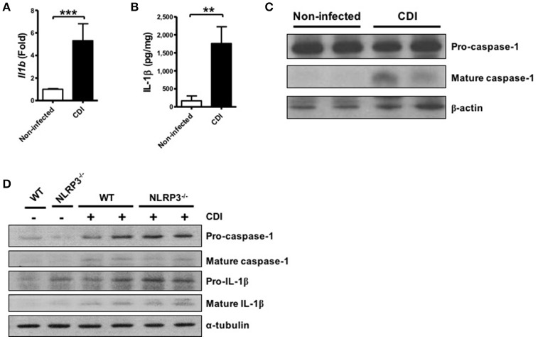
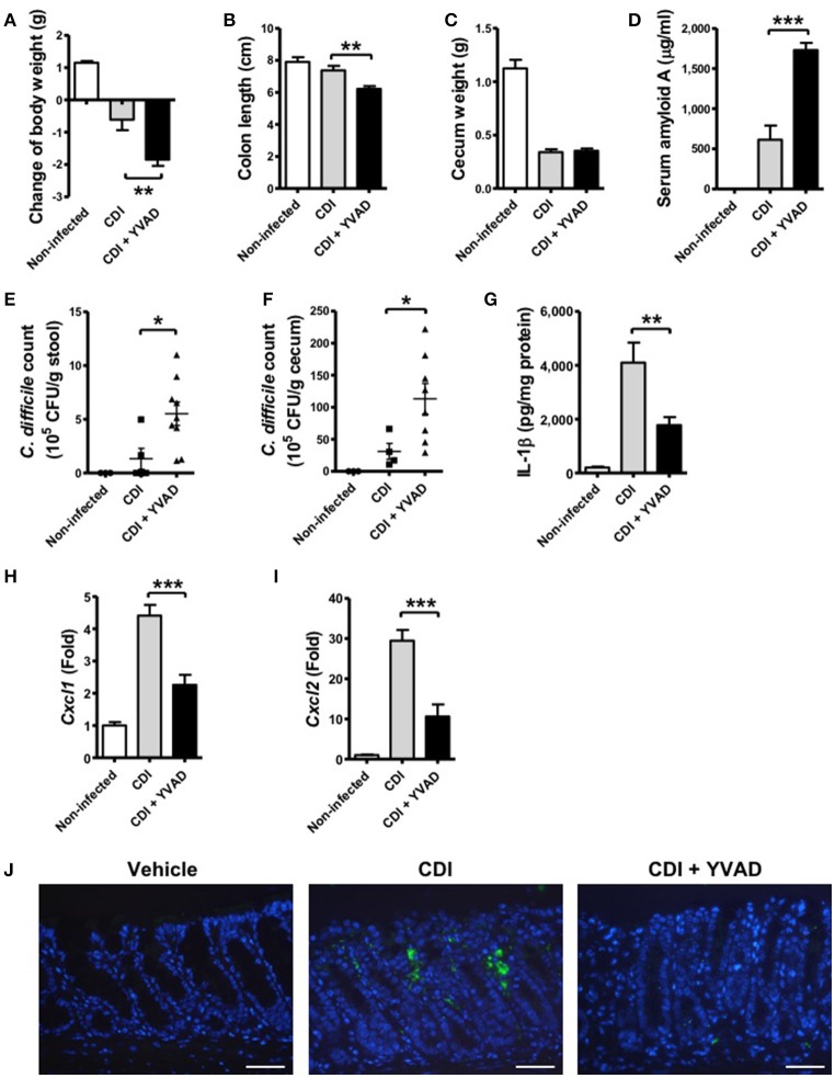
Clostridium difficile infection (CDI) is the leading cause of nosocomial infection in hospitalized patients receiving long-term antibiotic treatment. An excessive host inflammatory response is believed to be the major mechanism underlying the pathogenesis of C. difficile infection, and various proinflammatory cytokines such as IL-1β are detected in patients with C. difficile infection. IL-1β is known to be processed by caspase-1, a cysteine protease that is regulated by a protein complex called the inflammasome, which leads to a specialized form of cell death called pyroptosis. The function of inflammasome activation-induced pyroptosis is to clear or limit the spread of invading pathogens via infiltrated neutrophils. Here, we focused on inflammasome activation induced by intact C. difficile to re-evaluate the nature of inflammasome activation in CDI pathogenesis, which could provide information that leads to an alternative therapeutic strategy for the treatment of this condition in humans. First, we found that caspase-1-dependent IL-1β production was induced by C. difficile pathogens in macrophages and increased in a time-dependent manner. Moreover, intracellular toxigenic C. difficile was essential for ATP-P2X7 pathway of inflammasome activation and subsequent caspase-1-dependent pyroptotic cell death, leading to the loss of membrane integrity and release of intracellular contents such as LDH. Notably, we also observed that bacterial components such as surface layer proteins (SLPs) were released from pyroptotic cells. In addition, pro-IL-1β production was completely MyD88 and partially TLR2 dependent. Finally, to investigate the role of the caspase-1-dependent inflammasome in host defense, we found that colonic inflammasome activation was also induced by CDI and that caspase-1 inhibition by Ac-YVAD-CMK led to increased disease progression and C. difficile load. Taken together, the present results suggest that MyD88 and TLR2 are critical component in pro-IL-1β production and intracellular C. difficile following the ATP-P2X7 pathway of inflammasome activation and pyroptosis, which play important roles in host defense through the utilization of inflammation-mediated bacterial clearance mechanisms during C. difficile infection.

Keywords: ATP-P2X7 pathway; Clostridium difficile; MyD88; inflammasome activation; pyroptosis.


Figures

Figure 1
Figure 1
C. difficile induces caspase-1-dependent IL-1β production. (A,B) Peritoneal macrophages were infected with C. difficile VPI 10463 at increasing MOIs and a time course of infection was evaluated. Mature IL-1β and caspase-1 processing were analyzed by Western blotting. Lower panels showed quantification of mature caspase-1 and IL-1β relative protein level compared with non-infected group. (C) IL-1β and IL-18 production in the supernatant of infected cells were measured by ELISA. (D) Influence of the caspase-1 inhibitor Ac-YVAD-cmk (YVAD, 100 μM) on IL-1β release in infected macrophages by Western blot analysis. (E) The levels of secreted of IL-1β, IL-18, and TNF-α were determined by ELISA after treatment with YVAD. Values represent the mean ± SEM (N = 3/group). ***p < 0.001. N.D., not detected.
Figure 2
Figure 2
ATP-P2X7 pathway is essential for C. difficile-induced inflammasome activation. (A) ATP quantification in peritoneal macrophage supernatant after C. difficile VPI 10463 infection. (B–D) Peritoneal macrophages were infected with C. difficile VPI 10463 in the presence of apyrase (5 units/ml), glyburide (100 μM) or a 30-min pretreatment with the P2X7 antagonist (A438079) for 6 h. ***p < 0.001. Inflammasome activation, including IL-1β and caspase-1 processing, was analyzed by Western blotting. (E) The levels of IL-1β and IL-18 in the supernatant of infected cells after inhibitor treatment were determined by ELISA. ***p < 0.001, compared with vehicle-treated infected group. (F) Western blot analysis of inflammasome activation after transfection with scrambled siRNA or P2X7 siRNA in infected THP-1 cells. (G) IL-1β production and caspase-1 activation between WT and NLRP3−/− infected cells were detected by Western blotting. (H) The secretion of IL-1β, IL-18, and TNF-α were monitored by ELISA. Values represent the mean ± SEM (N = 3/group). ***p < 0.001. N.D., not detected. (I) Western blot analysis of inflammasome activation after transfection with scrambled siRNA or Pyrin siRNA in infected THP-1 cells.
Figure 3
Figure 3
Intracellular bacterial induces robust inflammasome activation and pyroptosis during CDI. (A) Peritoneal macrophages were infected with live or heat-killed C. difficile VPI 10463 for 6 h. Mature IL-1β and caspase-1 processing were analyzed by Western blotting. (B) The macrophages were infected with C. difficile in the presence of cytochalasin D (1 μM) for 6 h. Mature IL-1β and caspase-1 processing were analyzed by Western blotting. Peritoneal macrophages were infected with different strains of C. difficile (CCUG 37780, VPI 10463, and BAA 1805) for 6 h. (C) Pro-IL-1β and SLPs in cell lysate, and mature IL-1β, SLPs, and HMGB-1 in the supernatant of infected-macrophages were analyzed by Western blotting. (D) IL-1β production were assessed by ELISA. (E) Cell death induced by the different strains resulting in loss of membrane integrity was determined by the LDH release assay. (F) Mature IL-1β, SLPs, and HMGB-1 in the supernatant of infected-macrophages after caspase-1 inhibitor YVAD treatment were analyzed by Western blotting. (G) The LDH assay was also applied after the caspase-1 inhibitor Ac-YVAD-cmk (YVAD) treatment. Values represent the mean ± SEM (N = 3/group). *p < 0.05; ***p < 0.001. N.D., not detected.
Figure 4
Figure 4
C. difficile-induced pro-IL-1β production is completely MyD88 and partially TLR2 dependent. Peritoneal macrophages isolated from WT, MyD88−/−, Tlr2−/−, Tlr4−/−, and Tlr2−/−/Tlr4−/− mice were infected with C. difficile VPI 10463 for 6 h. (A) The mRNA gene expression levels of Il1b were determined by real-time PCR. (B) Pro-IL-1β production was analyzed in the cell lysate protein by Western blot analysis. (C) IL-1β production in the cell culture supernatant was assessed by ELISA. Values represent the mean ± SEM (N = 3/group). ***p < 0.001. N.D., not detected.
Figure 5
Figure 5
C. difficile infection induces colonic inflammasome activation. WT C57BL/6 mice were infected with C. difficile VPI 10463 for 2 days, and infected colon tissue was harvested for further analysis. (A) The mRNA gene expression levels of Il1b in the infected colon tissue were measured by real-time PCR. (B) IL-1β production in infected colon tissue was detected by ELISA. (C) Caspase-1 activation in the infected colon tissue was analyzed by Western blot. Values represent the mean ± SEM (N = 15/group). **p < 0.01; ***p < 0.001. (D) Mature caspase-1 and IL-1β production in the colon tissue of WT and NLRP3−/− mice with VPI 10463 infection were analyzed by Western blotting.
Figure 6
Figure 6
Inhibition of caspase-1-dependent inflammasome activation results in more severe disease progression during CDI. Mice were challenged with C. difficile VPI 10463 alone or together with an intraperitoneal injection of Ac-YVAD-cmk (12.5 μM), which is an irreversible caspase-1 inhibitor, for 2 days. Disease progression, including changes in (A) body weight, (B) colon length, and (C) cecum weight were monitored after 2 days of infection. (D) Serum amyloid A, which is an indicator of colitis, was measured by ELISA. The bacterial load of C. difficile in the feces (E) and cecum contents (F) were determined by plating on selective CCFA agar plates. (G) IL-1β production in the colon tissue was detected by ELISA. CXCL1 (H) and CXCL2 (I) mRNA levels in the colon tissue were analyzed by real-time PCR. Values represent the mean ± SEM (N = 5/group). *p < 0.05; **p < 0.01, ***p < 0.001. (J) Gr-1 positive cells in the colon tissue. Scale bar, 40 μM.

References

    1. Ali S. R., Timmer A. M., Bilgrami S., Park E. J., Eckmann L., Nizet V., et al. (2011). Anthrax toxin induces macrophage death by p38 MAPK inhibition but leads to inflammasome activation via ATP leakage. Immunity 35, 34–44. 10.1016/j.immuni.2011.04.015 - DOI - PMC - PubMed
    1. Altmeier S., Toska A., Sparber F., Teijeira A., Halin C., Leibundgut-Landmann S. (2016). IL-1 coordinates the neutrophil response to C. albicans in the oral mucosa. PLoS Pathog 12:e1005882. 10.1371/journal.ppat.1005882 - DOI - PMC - PubMed
    1. Ashida H., Mimuro H., Ogawa M., Kobayashi T., Sanada T., Kim M., et al. (2011). Cell death and infection: a double-edged sword for host and pathogen survival. J. Cell Biol. 195, 931–942. 10.1083/jcb.201108081 - DOI - PMC - PubMed
    1. Ausiello C. M., Cerquetti M., Fedele G., Spensieri F., Palazzo R., Nasso M., et al. (2006). Surface layer proteins from Clostridium difficile induce inflammatory and regulatory cytokines in human monocytes and dendritic cells. Microbes Infect. 8, 2640–2646. 10.1016/j.micinf.2006.07.009 - DOI - PubMed
    1. Bergsbaken T., Fink S. L., Cookson B. T. (2009). Pyroptosis: host cell death and inflammation. Nat. Rev. Microbiol. 7, 99–109. 10.1038/nrmicro2070 - DOI - PMC - PubMed

Publication types

MeSH terms