Review
. 2018 Jun;9(3):325-335.
doi: 10.1007/s13244-018-0611-8. Epub 2018 Apr 3.

How to use the Kaiser score as a clinical decision rule for diagnosis in multiparametric breast MRI: a pictorial essay

Affiliations
Review

How to use the Kaiser score as a clinical decision rule for diagnosis in multiparametric breast MRI: a pictorial essay

Matthias Dietzel et al. Insights Imaging. 2018 Jun.

Abstract

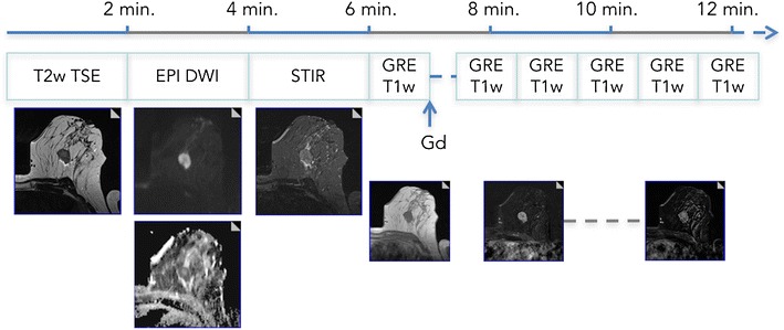
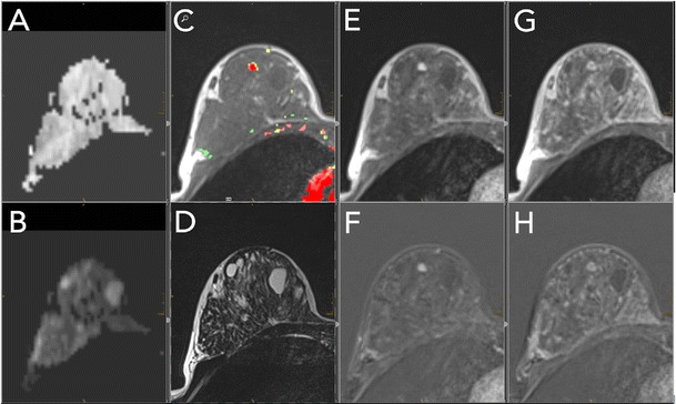
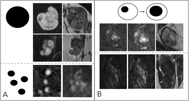
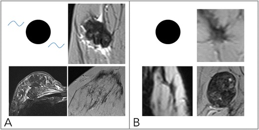
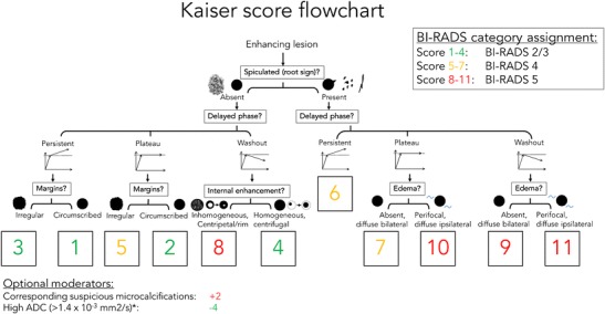
Due to its superior sensitivity, breast MRI (bMRI) has been established as an important additional diagnostic tool in the breast clinic and is used for screening in patients with an elevated risk for breast cancer. Breast MRI, however, is a complex tool, providing multiple images containing several contrasts. Thus, reading bMRI requires a structured approach. A lack of structure will increase the rate of false-positive findings and sacrifice most of the advantages of bMRI as additional work-up will be required. While the BI-RADS (Breast Imaging Reporting And Data System) lexicon is a major step toward standardised and structured reporting, it does not provide a clinical decision rule with which to guide diagnostic decisions. Such a clinical decision rule, however, is provided by the Kaiser score, which combines five independent diagnostic BI-RADS lexicon criteria (margins, SI-time curve type, internal enhancement and presence of oedema) in an intuitive flowchart. The resulting score provides probabilities of malignancy that can be used for evidence-based decision-making in the breast clinic. Notably, considerable benefits have been demonstrated for radiologists with initial and intermediate experience in bMRI. This pictorial essay is a practical guide to the application of the Kaiser score in the interpretation of breast MRI examinations.

Teaching points: • bMRI requires standardisation of patient-management, protocols, and reading set-up. • Reading bMRI includes the assessment of breast parenchyma, associated findings, and lesions. • Diagnostic decisions should be made according to evidence-based clinical decision rules. • The evidence-based Kaiser score is applicable independent of bMRI protocol and scanner. • The Kaiser score provides high diagnostic accuracy with low inter-observer variability.

Keywords: Breast MRI; Breast cancer; Clinical decision-making; Patient management.


Figures

Fig. 1
Fig. 1
Multiparametric bMRI protocol. A full triparametric bMRI protocol can be acquired in 15 min of magnet time. Patient preparation, including venous catheter placement, should be done outside the scanner room. The (recommended) axial protocol starts with precontrast T2w-TSE, DWI, and STIR (optional) sequences. Here, a mass lesion in a 47-year-old patient with perifocal oedema and central necrosis is easily depicted. Dynamic scanning should include 4–5 min of post-contrast acquisition. The example demonstrates a type III curve, and the resulting Kaiser score is 8 (see Fig. 6), highly suggestive of malignancy. Histopathology revealed an invasive ductal cancer G3
Fig. 2
Fig. 2
Standardised hanging protocol/reading layout for a two-screen solution. Standardised hanging protocols decrease the reading time and increase diagnostic confidence while reducing potential sources of errors. All series should be linked, allowing simultaneous scrolling and zooming. This example shows a 44-year-old high-risk patient. A: ADC map; B: high b-value DWI image; C: pre-contrast T1w (here with superimposed colour-coded enhancement map indicating dominant wash-out, coded in red); D: T2w-TSE; E/F: early contrast-enhanced scan with corresponding subtraction; G/H: delayed contrast-enhanced scan with corresponding subtraction within the dynamic series (E–H); the same windows and centre setting should be used. This step should be performed for subtracted and non-subtracted images separately. An incidental lesion of 5 mm in size is present in the right breast, showing homogeneous enhancement (E, F), followed by early and central wash-out (C, G, H). Intermediate T2w-signal [D, no restricted diffusivity (A, B)]. The lesion corresponds to a Kaiser score of 4 (see Fig. 6); MRI-guided biopsy revealed a benign adenosis B2
Fig. 3
Fig. 3
Diagnostic criteria: margins. Margins can be either circumscribed or not circumscribed. Circumscribed margins indicate a benign lesion and are more regularly found in mass (A) than in non-mass (B) lesions. Non-circumscribed margins include irregular (C: mass lesion, D: non-mass lesion), hinting at an intermediate risk of breast cancer, and spiculated. Spiculated margins (E: mass lesions, F: non-mass lesion) are highly suggestive of malignancy. Note that the most suspicious criterion applies; thus, single spiculae in an otherwise circumscribed lesion constitute spiculated margins. This is why this criterion was named the “root sign” by Kaiser
Fig. 4
Fig. 4
Diagnostic criteria: non-suspicious internal enhancement patterns. A: Homogeneous enhancement suggests benign lesions. Note that even a homogeneous enhancing lesion may show areas of lesser or absent enhancement due to septae and fibrotic parts [A, upper two rows, each early enhanced subtractions (left) and T2w (right)]. Homogeneity is more difficult to assess in non-mass lesions (A, lower row) and includes homogeneous internal morphology. Therefore, this feature was referred to as “stippled” enhancement in the initial BI-RADS lexicon. B: A central or centrifugal enhancement is highly suggestive of a benign lesion. To assess this feature, pre-contrast images need to be considered. While this feature usually applies to mass lesions only (A, upper row, from right to left, early, delayed enhanced, and T2w images), non-mass lesions may also present with this typical benign feature (B, lower row, note the by far larger lesion correlate on the right T2w image, as marked by arrows)
Fig. 5
Fig. 5
Diagnostic criteria: suspicious internal enhancement patterns. Heterogeneous enhancement may be associated with breast cancer and applies to mass and non-mass lesions (A, upper and lower row, respectively). Specific for malignancy is a centripetal or rim-like enhancement (B). In particular, a broad heterogeneous rim, regularly associated with a delayed enhancement of the central lesion parts, is highly suggestive of breast cancer (B, upper row). However, a thin, rather subtle and homogeneous rim enhancement with absent enhancement of the lesion centre hints at inflammatory conditions, such as cysts, liponecrosis, and abscess (B, upper row, left upper lesion). In non-mass lesions, rim enhancement may appear in a multiple and clustered manner (B, lower row) and is referred to as “clustered ring enhancement” in the BI-RADS lexicon
Fig. 6
Fig. 6
Diagnostic criteria: oedema. As shown in (A), oedema is present if fluid-caused T2w hyperintensity not attributable to cysts or dilated ducts is identified in the breast that harbours an enhancing lesion. Odema may be perifocal [A, upper right (mass) and lower right (non-mass)] or more diffuse (A, lower left, showing subcutaneous, diffuse, and pre-/intrapectoral oedema in an inflammatory breast cancer case with distinct lymphangiosis). B: Absent oedema. The common pitfall causing false-positive oedema assessment is chemical shift artefacts in sharp fat-water interfaces, such as that caused by vessels (e.g., B upper right, lower right) and capsulated fibroadenomas at the parenchyma-fat interface
Fig. 7
Fig. 7
The Kaiser score flowchart. The Kaiser score is assigned by following a simple flowchart from the top to the bottom, which lets the reader assign the presence or absence of four diagnostic criteria (presence of spiculation being a formal subgroup of margin assessment according to BI-RADS). The resulting score is associated with an increasing risk of malignancy (from 1 to 11) and can be translated into BI-RADS categories as follows: 1–4: minimal risk of breast cancer—BI-RADS 2/3; 5–7: intermediate risk of breast cancer—BI-RADS 4; 8–11: high risk of breast cancer—BI-RADS 5. Optional moderators include: presence of suspicious corresponding microcalcifications (+2) and high corresponding ADC values (-4). Further details and explanations are provided in the main text
Fig. 8
Fig. 8
Early (A) and delayed (B) contrast-enhanced subtractions, T2w (C), and ADC map (D). Mass lesion in the left breast of a 27-year-old female, newly palpable after considerable weight loss. The lesion shows no spiculations, a persistent enhancement curve type, and circumscribed margins, corresponding to a Kaiser score of 1 (Fig. 6). Note internal septations and high ADC (2.1*10-3 mm2/s). This is an unambiguous benign finding, BI-RADS 2. The patient requested a biopsy that revealed a fibroadenoma B2
Fig. 9
Fig. 9
Early (A) and delayed (B) contrast-enhanced subtractions, T2w (C), and ADC map (D). Mass lesion in the left breast of a 41-year-old female with newly developed bloody discharge. The lesion shows no spiculations, circumscribed margins (first subtraction), and a plateau-type enhancement curve, corresponding to a Kaiser score of 2, practically excluding malignancy (Fig. 6). Note the intraductal lesion location on T2w and the intermediate low ADC (1.1*10-3 mm2/s). Findings are pathognomonic for a benign papilloma. The symptomatic lesion was resected and histopathology revealed a benign papilloma without atypia
Fig. 10
Fig. 10
Early (A) and delayed (B) contrast-enhanced subtractions, T2w (C), and ADC map (D). Non-mass lesion in the left breast of a 25-year-old female with pain after stopping breast-feeding and unclear sonographic findings. The lesion shows no spiculations, a persistent enhancement curve type, and irregular margins, corresponding to a Kaiser score of 3 (Fig. 6). Note the internal small cystic correlate that suggests a benign lesion; ADC is unspecific (1.2*10-3 mm2/s). Together with the clinical symptoms, periductal mastitis must be suspected and it is safe to assign BI-RADS 2. Due to personal preferences, the patient requested a biopsy that revealed chronic periductal mastitis, B2
Fig. 11
Fig. 11
Early (A) and delayed (B) contrast-enhanced subtractions, T2w (C), and ADC map (D). Mass lesion in the right breast of a 50-year-old female undergoing follow-up for breast cancer treatment. The lesion shows spiculations and a persistent enhancement curve type, corresponding to a Kaiser score of 6 (Fig. 6). T2w signal intensity is low (C) and ADC is decreased (0.8*10-3 mm2/s). Histopathology revealed recurrent invasive ductal cancer G2, B5b
Fig. 12
Fig. 12
Early (A) and delayed (B) contrast-enhanced subtractions, T2w (C), and ADC map (D). Mass and non-mass lesions in the left breast of a 48-year-old female undergoing MRI for staging purposes because of conventional BI-RADS 5 findings prior to biopsy. The main mass lesion shows multiple subtle spiculations, a wash-out curve type, and a perifocal oedema, corresponding to a Kaiser score of 11 (Fig. 6). Note central necrosis (high signal in C) and low ADC (0.7*10-3 mm2/s). The anterior lesions show no spiculations, a plateau curve type, and irregular margins, corresponding to a Kaiser score of 5 (Fig. 6). Again, ADC is low (0.9-1*10-3 mm2/s). Histopathology revealed a multicentric IDC, G3, with a DCIS component, B5b
Fig. 13
Fig. 13
Early (A) and delayed (B) contrast-enhanced subtractions, T2w (C), and ADC map (D). Mass lesion in the left breast of a 50-year-old female with several unclear mass lesions in both breasts; MRI obtained for treatment planning. The lesion shows no spiculations, a wash-out enhancement curve type, and a heterogeneous internal enhancement, corresponding to a Kaiser score of 8 (Fig. 6). Note high signal intensity on T2w. Due to high ADC (1.9*10-3 mm2/s), the lesion was downgraded to a Kaiser score of 4. Histopathology revealed a fibroadenoma with regressive changes, B2

References

    1. Kaiser WA, Zeitler E. MR imaging of the breast: fast imaging sequences with and without Gd-DTPA. Preliminary observations. Radiology. 1989;170:681–686. doi: 10.1148/radiology.170.3.2916021. - DOI - PubMed
    1. Heywang SH, Hilbertz T, Pruss E, et al. Dynamic contrast medium studies with flash sequences in nuclear magnetic resonance tomography of the breast. Digitale Bilddiagn. 1988;8:7–13. - PubMed
    1. Warner E, Messersmith H, Causer P, et al. Systematic review: using magnetic resonance imaging to screen women at high risk for breast cancer. Ann Intern Med. 2008;148:671–679. doi: 10.7326/0003-4819-148-9-200805060-00007. - DOI - PubMed
    1. Mann RM, Kuhl CK, Kinkel K, Boetes C. Breast MRI: guidelines from the European Society of Breast Imaging. Eur Radiol. 2008;18:1307–1318. doi: 10.1007/s00330-008-0863-7. - DOI - PMC - PubMed
    1. Sardanelli F, Boetes C, Borisch B, et al. (2010) Magnetic resonance imaging of the breast: recommendations from the EUSOMA working group. Eur J Cancer Oxf Engl. 1990;46:1296–1316. doi: 10.1016/j.ejca.2010.02.015. - DOI - PubMed

LinkOut - more resources