. 2018 May 18;293(20):7727-7736.
doi: 10.1074/jbc.RA117.000600. Epub 2018 Apr 4.

The full-length cytochrome P450 enzyme CYP102A1 dimerizes at its reductase domains and has flexible heme domains for efficient catalysis

Affiliations

The full-length cytochrome P450 enzyme CYP102A1 dimerizes at its reductase domains and has flexible heme domains for efficient catalysis

Haoming Zhang et al. J Biol Chem. .

Abstract

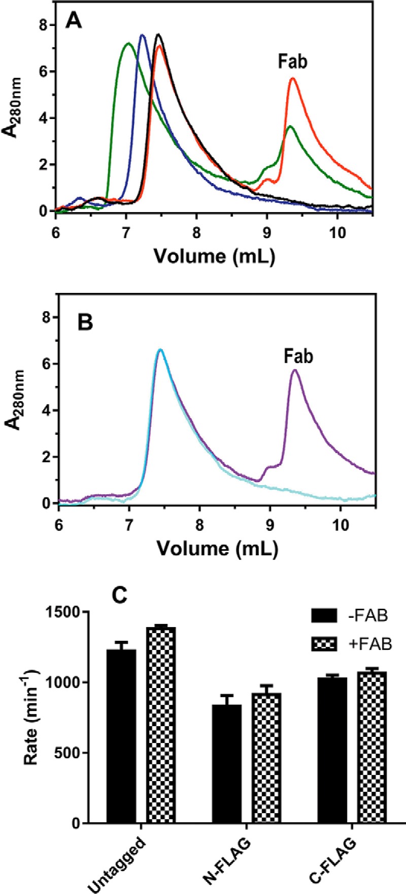
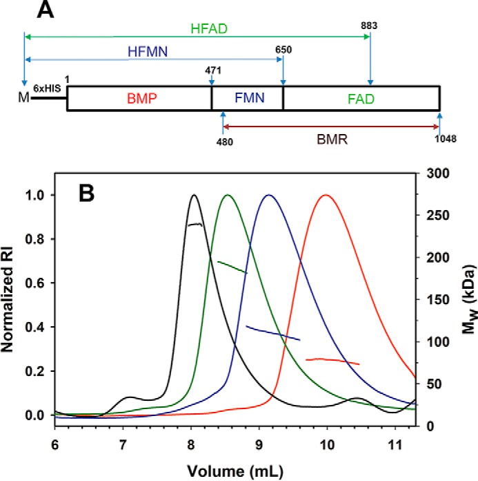
The cytochrome P450 enzyme CYP102A1 from Bacillus megaterium is a highly efficient hydroxylase of fatty acids, and there is a significant interest in using CYP102A1 for biotechnological applications. Here, we used size-exclusion chromatography-multiangle light scattering (SEC-MALS) analysis and negative-stain EM to investigate the molecular architecture of CYP102A1. The SEC-MALS analysis yielded a homogeneous peak with an average molecular mass of 235 ± 5 kDa, consistent with homodimeric CYP102A1. The negative-stain EM of dimeric CYP102A1 revealed four distinct lobes, representing the two heme and two reductase domains. Two of the lobes were in close contact, whereas the other two were often observed apart and at the ends of a U-shaped configuration. The overall dimension of the dimer was ∼130 Å. To determine the identity of the lobes, we FLAG-tagged the N or C terminus of CYP102A1 to visualize additional densities in EM and found that anti-FLAG Fab could bind only the N-tagged P450. Single-particle analysis of this anti-Flag Fab-CYP102A1 complex revealed additional density in the N-terminally tagged heme domains, indicating that the heme domains appear flexible, whereas the reductase domains remain tightly associated. The effects of truncation on CYP102A1 dimerization, identification of cross-linked sites by peptide mapping, and molecular modeling results all were consistent with the dimerization of the reductase domain. We conclude that functional CYP102A1 is a compact globular protein dimerized at its reductase domains, with its heme domains exhibiting multiple conformations that likely contribute to the highly efficient catalysis of CYP102A1.

Keywords: cytochrome P450; cytochrome P450 BM3; dimerization; electron microscopy (EM); electron transfer; fatty acid hydroxylase; heme domain; protein structure; reductase domain.


Conflict of interest statement

The authors declare that they have no conflicts of interest with the contents of this article

Figures

Figure 1.
Figure 1.
Analysis of full-length CYP102A1 by SEC–MALS and negative stain EM. A, SEC–MALS trace of CYP102A1. CYP102A1 was eluted from a WTC-050S5 column (7.8 × 300 mm, 5 μm, Wyatt Technology) as a dimeric complex peak at ∼8.5 ml. B, high contrast micrographs of CYP102A1 dimer. C, reference free 2D class averages using Relion 2D classification show four distinct densities and multiple conformations of dimeric CYP102A1.
Figure 2.
Figure 2.
SEC analysis and catalytic activity of control, N-FLAG, and C-FLAG CYP102A1. To form CYP102A–Fab complexes, CYP102A1 (10 μm) was preincubated with 20 μm Fab in PBS buffer at 4 °C overnight. The incubation was then diluted by 5-fold for SEC analysis and by 100-fold for determination of the rate of NADPH consumption. The rates of NADPH consumption were determined spectrophotometrically by observing time-dependent decrease in absorbance at 340 nm after the addition of 0.2 mm NADPH to CYP102A1 as described under “Experimental procedures.” A, SEC traces of the untagged and N-FLAG-tagged CYP102A1 in the presence and absence of Fab. Aliquots of ∼100 μl of the incubation were loaded onto a Yarra SEC-3000 column (7.8 × 300 mm, 5 μm, Phenomenex) and separated in PBS buffer at 0.4 ml/min. Black trace, untagged CYP102A1; red trace, untagged CYP102A1 + Fab; blue trace, N-FLAG CYP102A1; green trace, N-FLAG CYP102A1 + Fab. B, SEC traces of the C-FLAG-tagged CYP102A1 showing absence of CYP102A1–Fab complex. Cyan trace, C-FLAG CYP102A1; purple trace, C-FLAG CYP102A1 + Fab. C, rates of NADPH consumption in the presence and absence of Fab.
Figure 3.
Figure 3.
Analysis of N-FLAG CYP102A1–Fab complex by SEC–MALS and negative stain EM. A, SEC–MALS trace of N-FLAG CYP102A1 in the presence and absence of Fab. To form the N-FLAG CYP102A–Fab complex, N-FLAG CYP102A1 was preincubated with 2-fold excess Fab at 4 °C overnight. The protein samples were then eluted at 0.4 ml/min from a Yarra SEC-3000 column. B, single particles of dimeric N-FLAG CYP102A1–Fab complex in the U-shaped 2D class average. The white arrows indicate the density for Fab. The U-shaped 2D class average is identical to that shown in Fig. 1C. RI, refractive index.
Figure 4.
Figure 4.
SEC–MALS analysis of truncated forms of CYP102A1. A, schematic representation of truncated CYP102A1 construct BMP, HFMN, HFAD, and BMR used in this study. B, SEC–MALS analysis of the truncated constructs of CYP102A1. Truncated CYP102A1 (∼8 μm) was eluted from a WTC-050S5 column at 0.4 ml/min in PBS buffer. Green trace, HFAD; blue trace, HFMN; red trace, BMP. The control is full-length CYP102A1 shown in black. The left y axis is normalized intensity of refractive index (RI), whereas the right y axis is molecular mass determined by static MALS as described under “Experimental procedures.” Molecular mass is shown as a straight line in a color corresponding to the SEC–MALS trace.
Figure 5.
Figure 5.
Analysis of cross-linked full-length CYP102A1 (FL), BMP, and BMR domains by SDS–PAGE. Protein samples (∼8 μm) were cross-linked by DSS at 0, 5, 50, and 500 μm and quenched with 50 mm ammonium bicarbonate as described under “Experimental procedures.” Aliquots of the cross-linked samples were mixed with equal volumes of Laemmli buffer and denatured at 98 °C for 5 min. The denatured samples were separated on 4–20% gradient Tris/glycein gels and stained with Coomassie Blue for visualization.
Figure 6.
Figure 6.
3D model of dimeric CYP102A1 showing the close view of the dimerization interface. The 3D EM density shown in solid surface was constructed from the 2D class average images as described under “Experimental procedures.” The crystal structure of the BMP (PDB code 4KEW) and the BMR homology model were fit to the 3D density volume using Chimera. The BMR homology model was constructed with Modeler 9v8 as described previously (39). BMP molecule is shown in magenta, and the FMN, FAD, and nucleotide-binding domains of the BMR are shown in green, blue, and cyan, respectively. Co-factor FMN, FAD, and heme are shown in orange, yellow, and red, respectively. Residues Lys777 and Lys790 are shown in brown and gray, respectively.
Scheme 1.
Scheme 1.
Schematic representation of a dimeric model of CYP102A1.

References

    1. Narhi L. O., and Fulco A. J. (1986) Characterization of a catalytically self-sufficient 119,000-dalton cytochrome P-450 monooxygenase induced by barbiturates in Bacillus megaterium. J. Biol. Chem. 261, 7160–7169 - PubMed
    1. Noble M. A., Miles C. S., Chapman S. K., Lysek D. A., MacKay A. C., Reid G. A., Hanzlik R. P., and Munro A. W. (1999) Roles of key active-site residues in flavocytochrome P450 BM3. Biochem. J. 339, 371–379 10.1042/0264-6021:3390371,10.1042/bj3390371 - DOI - PMC - PubMed
    1. Truan G., and Peterson J. A. (1998) Thr268 in substrate binding and catalysis in P450BM-3. Arch. Biochem. Biophys. 349, 53–64 10.1006/abbi.1997.0400 - DOI - PubMed
    1. Venkataraman H., Te Poele E. M., Rosloniec K. Z., Vermeulen N., Commandeur J. N., van der Geize R., and Dijkhuizen L. (2015) Biosynthesis of a steroid metabolite by an engineered Rhodococcus erythropolis strain expressing a mutant cytochrome P450 BM3 enzyme. Appl. Microbiol. Biotechnol. 99, 4713–4721 - PubMed
    1. Kim K. H., Kang J. Y., Kim D. H., Park S. H., Park S. H., Kim D., Park K. D., Lee Y. J., Jung H. C., Pan J. G., Ahn T., and Yun C. H. (2011) Generation of human chiral metabolites of simvastatin and lovastatin by bacterial CYP102A1 mutants. Drug Metab. Dispos. 39, 140–150 10.1124/dmd.110.036392 - DOI - PubMed

Publication types

MeSH terms

LinkOut - more resources