Review
. 2018 Apr 4;31(3):e00084-16.
doi: 10.1128/CMR.00084-16. Print 2018 Jul.

Options and Limitations in Clinical Investigation of Bacterial Biofilms

Affiliations
Review

Options and Limitations in Clinical Investigation of Bacterial Biofilms

Maria Magana et al. Clin Microbiol Rev. .

Abstract

Bacteria can form single- and multispecies biofilms exhibiting diverse features based upon the microbial composition of their community and microenvironment. The study of bacterial biofilm development has received great interest in the past 20 years and is motivated by the elegant complexity characteristic of these multicellular communities and their role in infectious diseases. Biofilms can thrive on virtually any surface and can be beneficial or detrimental based upon the community's interplay and the surface. Advances in the understanding of structural and functional variations and the roles that biofilms play in disease and host-pathogen interactions have been addressed through comprehensive literature searches. In this review article, a synopsis of the methodological landscape of biofilm analysis is provided, including an evaluation of the current trends in methodological research. We deem this worthwhile because a keyword-oriented bibliographical search reveals that less than 5% of the biofilm literature is devoted to methodology. In this report, we (i) summarize current methodologies for biofilm characterization, monitoring, and quantification; (ii) discuss advances in the discovery of effective imaging and sensing tools and modalities; (iii) provide an overview of tailored animal models that assess features of biofilm infections; and (iv) make recommendations defining the most appropriate methodological tools for clinical settings.

Keywords: animal host models; biofilms; flow cells; imaging; quantification.


Figures

FIG 1
FIG 1
Developmental stages in biofilm formation. One or more planktonic bacterial species adhere to a biotic/abiotic surface. Attached bacteria grow as a multicellular community, forming microcolonies in which they multiply and mature. This microbial infrastructure results in the development of a mature biofilm. Eventually, biofilms serve as bacterial reservoirs that are transmitted back to the environment through biofilm dispersal and then colonize new surfaces. (The concept of this figure was inspired by reference .)
FIG 2
FIG 2
Biofilm types. (A) Surface-attached biofilms form colonies on a solid surface and are highly dependent on the substratum material. (B) Pellicles are formed in the air-liquid interface of fluids in nature or in the lab. Cells are bound together, forming a distinct macroscopic floating infrastructure. Thick pellicle formation requires the presence of exopolysaccharides (EPS). (C) Submerged biofilms develop under flow conditions. Biofilm formation under flow conditions is achieved in either indwelling catheters or suitably adapted lab devices.
FIG 3
FIG 3
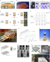
Laboratory setups. (A) Colony biofilms on agar plates. (1) Schematic diagram of a colony biofilm. (2) Various types of macrocolonies grown on agar medium. (Panel 1 reprinted from reference with permission of the publisher; panel 2 reprinted from reference .) (B) Microtiter plate. (Photograph taken and kindly provided by Alex Hall.) (C) Air-liquid biofilms. (1) Pellicle formation at the air-liquid surface. (2) Crystal violet staining was performed to assess air-liquid biofilm formation on abiotic surfaces. (Panel 1 reprinted from reference ; panel 2 reprinted from reference .) (D) BioFilm ring test. Photos of scanning microplates were taken with a plate reader after magnetization and show no biofilm formation (1) and biofilm formation (2). (Photos reprinted from reference .) (E) Kadouri biofilm system for flow biofilm study. (Reprinted from reference .) (F) Drip-flow reactor and various components. (Photograph kindly provided by the Center for Biofilm Engineering, MSU-Bozeman; see reference for further details.) (G) Modified Robbins device. (Reprinted from reference .) (H) Rotating-disk reactor. (Photograph kindly provided by BioSurface Technologies Corp.) (I) Peg lid Calgary device. (Adapted from reference with permission from Macmillan Publishers Ltd.) (J) Biofilm growth in microfermentors. (Reprinted from reference .) (K) Microfluidic device and experimental setup for biofilm formation (Reprinted from reference with permission.)
FIG 4
FIG 4
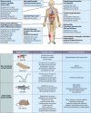
Comparative visualization of biofilm-attributed human infections and classes of major vertebrate and nonvertebrate models developed. This is not an extensive list, but presentation of the bacterial strains that are most commonly encountered in biofilm-related research studies is included.
FIG 5
FIG 5
Illustrative quantitative and time-dependent representation of biofilm-based methodology publications for currently used devices and assays. CV, crystal violet; DAPI, 4′,6-diamidino-2-phenylindole; CTC, oxidized 5-cyano-2,3-ditolyl tetrazolium chloride; AO, acridine orange; AB, alamarBlue; DMMB, dimethylmethylene blue; BTA, biotimer assay; FDA, fluorescein diacetate; XTT, 2,3-bis(2-methoxy-4-nitro-5-sulfophenyl)-5-[(phenylamino)carbonyl]-2H-tetrazolium hydroxide; CR, Congo red.
None
None
None
None
None
None
None
None
None
None

References

    1. Wimpenny J, Manz W, Szewzyk U. 2000. Heterogeneity in biofilms. FEMS Microbiol Rev 24:661–671. doi:10.1111/j.1574-6976.2000.tb00565.x. - DOI - PubMed
    1. Monds RD, O'Toole GA. 2009. The developmental model of microbial biofilms: ten years of a paradigm up for review. Trends Microbiol 17:73–87. doi:10.1016/j.tim.2008.11.001. - DOI - PubMed
    1. Stoodley P, Sauer K, Davies DG, Costerton JW. 2002. Biofilms as complex differentiated communities. Annu Rev Microbiol 56:187–209. doi:10.1146/annurev.micro.56.012302.160705. - DOI - PubMed
    1. O'Toole G, Kaplan HB, Kolter R. 2000. Biofilm formation as microbial development. Annu Rev Microbiol 54:49–79. doi:10.1146/annurev.micro.54.1.49. - DOI - PubMed
    1. Lebeaux D, Chauhan A, Rendueles O, Beloin C. 2013. From in vitro to in vivo models of bacterial biofilm-related infections. Pathogens 2:288–356. doi:10.3390/pathogens2020288. - DOI - PMC - PubMed

Publication types