Review
. 2018 Apr;208(4):1311-1336.
doi: 10.1534/genetics.117.300157.

Wingless Signaling: A Genetic Journey from Morphogenesis to Metastasis

Affiliations
Review

Wingless Signaling: A Genetic Journey from Morphogenesis to Metastasis

Amy Bejsovec. Genetics. 2018 Apr.

Abstract

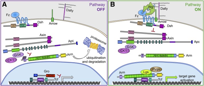
This FlyBook chapter summarizes the history and the current state of our understanding of the Wingless signaling pathway. Wingless, the fly homolog of the mammalian Wnt oncoproteins, plays a central role in pattern generation during development. Much of what we know about the pathway was learned from genetic and molecular experiments in Drosophila melanogaster, and the core pathway works the same way in vertebrates. Like most growth factor pathways, extracellular Wingless/Wnt binds to a cell surface complex to transduce signal across the plasma membrane, triggering a series of intracellular events that lead to transcriptional changes in the nucleus. Unlike most growth factor pathways, the intracellular events regulate the protein stability of a key effector molecule, in this case Armadillo/β-catenin. A number of mysteries remain about how the "destruction complex" destabilizes β-catenin and how this process is inactivated by the ligand-bound receptor complex, so this review of the field can only serve as a snapshot of the work in progress.

Keywords: FlyBook; Wingless; Wnt; beta-catenin; signal transduction.


Figures

Figure 1
Figure 1
Viable wg mutant phenotypes. (A) Normal bristle pattern on the notum, the back of a fly’s thorax, with both halteres visible out of focus at the posterior edge (in this and all images, posterior is to the right). (B) Notum of a wg1 homozygous mutant showing disrupted pattern and absence of both wings and one haltere. (C) Side view of wild-type (WT) fly. (D) Side view of wg1 homozygous mutant showing duplicated notum in place of one missing wing, and misshapen eye (cinnabar eye color is not part of the wg phenotype). (E) WT eye shows a regular pattern of ommatidia, the units of the compound eye. (F) The eye of a fly heterozygous for the Gla mutation shows a smooth “glazed” surface.
Figure 2
Figure 2
Generation of genetic mosaics. (A) Clones of homozygous mutant cells are generated in heterozygous flies when mitotic recombination between the homologs occurs. This rare event can be induced by exposing flies to X-rays, which cause double-stranded DNA breaks that often lead to crossing-over in the process of being repaired. If both mutant chromatids are pulled to the same mitotic spindle pole, the resulting two daughter cells will have different genotypes. Subsequent cell divisions generate mitotic clones from each daughter, producing a “twin spot” of homozygous mutant (red) and homozygous wild-type (WT) (blue) cells within a field of heterozygous cells. (B) The yeast flippase (FLP) and its target sequence (FRT) can be used instead of X-rays to induce mitotic recombination. Heat shock-induced flippase catalyzes site-specific recombination at the FRT target. The presence of a dominant marker, such as green fluorescent protein (GFP), on the WT homolog allows easy detection of mutant clones in somatic tissue. Inclusion of the ovoD dominant female-sterile mutation blocks egg formation in heterozygous ovarian tissue. During the production of germ line clones, the only eggs recovered are derived from tissue homozygous for the non-ovoD-bearing chromosome.
Figure 3
Figure 3
Embryonic wg phenotypes. (A) Wild-type (WT) embryos secrete a segmental pattern of denticle belts separated by naked cuticle on their ventral surface. Bar, 50 µm. (B) wg null mutant embryos produce a cuticle pattern with no naked cuticle, only denticles, on the ventral surface. (C) Wg antibody staining (red) shows that the protein is expressed in stripes in WT embryos, and the protein is detected over several cell diameters on either side of the stripe (cell outlines visualized with Neurotactin antibody staining, green). The stripes of wg expression are located within a subset of the epidermal cells that will secrete naked cuticle. (D) Arm antibody staining (white) in WT embryos shows higher levels of Arm in broad stripes that are roughly centered over the Wg-producing cells.
Figure 4
Figure 4
The magic of balancer chromosomes. (A) A lethal mutation must be maintained in the heterozygous state, with a wild-type allele of the gene on the other homolog. If the other homolog is entirely wild-type, this wild-type chromosome will predominate in future generations, and the lethal mutation may be lost. (B) Balancers, such as Curly-O (CyO), were designed so that homozygous lethal mutations could be maintained in heterozygous, balanced, fly stocks that are stable over many generations. Flies carrying a lethal mutation on one homolog and a balancer chromosome as the other homolog would produce progeny where one-quarter are homozygous lethal due to the first mutation, one-quarter are homozygous lethal (or are sterile, in the case of X chromosome balancers) due to homozygosity for the balancer, and the remaining half survive to adulthood as heterozygotes with a genotype identical to the parents. Multiple inversions on the balancer disrupt pairing between the homologs, preventing a recombination event between the desired lethal mutation and the recessive lethal mutation carried by the balancer, which could otherwise generate an entirely wild-type chromosome. (C) Balancer chromosomes can be used in genetic screens to isolate new lethal mutations. Flies are mutagenized and crossed to a stock carrying a balancer chromosome. The Heidelberg screens made use of a balancer stock carrying a dominant temperature-sensitive lethal mutation (here designated Let ts) to eliminate the nonbalancer chromosome in the next generation. However, any dominant mutation different from the marker on the balancer could be used and selected against in the next generation. Each F1 individual is then crossed back to a fly of the opposite sex from the balancer stock. F2 progeny from these individual crosses are then selected for the presence of the balancer dominant marker and the absence of the nonbalancer dominant marker, or incubated at high temperature to eliminate the nonbalancer progeny if using a dominant temperature-sensitive lethal stock. In this way, F2 individuals of the opposite sex, each heterozygous for the same mutagenized chromosome, are generated and can be crossed together to assess the homozygous phenotype of the mutagenized chromosome.
Figure 5
Figure 5
Diagram of core components in the Wg pathway. (A) In the absence of Wg signaling, Arm is presented by Apc and Axin to CK1 and Zw3/GSK for phosphorylation, targeting it for ubiquitination and degradation by the proteasome. Tcf binds target genes and, with the Gro transcriptional corepressor, keeps expression repressed. (B) Wg, concentrated at the cell surface by glycosaminoglycans on the glypican Dally, binds the Fz and Arrow receptors and causes them to cluster. This allows polymerization of Dsh and Axin at the plasma membrane, inactivating the kinase complex so that it cannot target Arm for destruction. Stabilized Arm translocates into the nucleus, binds to Tcf, and recruits the transcriptional activation complex, which includes Lgs and Pygo. Structural features of proteins depicted here are based on data from Wodarz and Nusse (1998), Janda et al. (2012).
Figure 6
Figure 6
Diagram of putative Wg protein structure. The structure of Xenopus Wnt8 was solved by cocrystallizing it with the mouse Fz8 CRD, allowing determination of the positions for glycosylations, conserved cysteines involved in disulfide bonds, and the fatty acyl attachment (Janda et al. 2012). Cylinders depict α-helical regions and block arrows depict β-sheets predicted in the crystal structure. Approximate positions of wg mutations are indicated by stars (those implicated in Wg protein transport are within the blue box), and the position of the nonconserved insert region of Wg is indicated in gray (van den Heuvel et al. 1993; Bejsovec and Wieschaus 1995; Dierick and Bejsovec 1998).

References

    1. Adler P. N., 1992. The genetic control of tissue polarity in Drosophila. Bioessays 14: 735–741. - PubMed
    1. Ahmed Y., Hayashi S., Levine A., Wieschaus E., 1998. Regulation of Armadillo by a Drosophila APC inhibits neuronal apoptosis during retinal development. Cell 93: 1171–1182. - PubMed
    1. Ahmed Y., Nouri A., Wieschaus E., 2002. Drosophila Apc1 and Apc2 regulate Wingless transduction throughout development. Development 129: 1751–1762. - PubMed
    1. Akong K., Grevengoed E. E., Price M. H., McCartney B. M., Hayden M. A., et al. , 2002. Drosophila APC2 and APC1 play overlapping roles in Wingless signaling in the embryo and imaginal discs. Dev. Biol. 250: 91–100. - PubMed
    1. Alexandre C., Lecourtois M., Vincent J., 1999. Wingless and Hedgehog pattern Drosophila denticle belts by regulating the production of short-range signals. Development 126: 5689–5698. - PubMed

Publication types

LinkOut - more resources