Review
. 2018 Apr;208(4):1337-1349.
doi: 10.1534/genetics.118.300755.

Biology in Bloom: A Primer on the Arabidopsis thaliana Model System

Affiliations
Review

Biology in Bloom: A Primer on the Arabidopsis thaliana Model System

Andrew W Woodward et al. Genetics. 2018 Apr.

Abstract

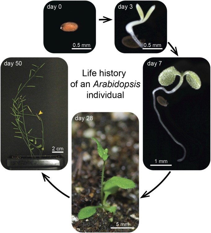
Arabidopsis thaliana could have easily escaped human scrutiny. Instead, Arabidopsis has become the most widely studied plant in modern biology despite its absence from the dinner table. Pairing diminutive stature and genome with prodigious resources and tools, Arabidopsis offers a window into the molecular, cellular, and developmental mechanisms underlying life as a multicellular photoautotroph. Many basic discoveries made using this plant have spawned new research areas, even beyond the verdant fields of plant biology. With a suite of resources and tools unmatched among plants and rivaling other model systems, Arabidopsis research continues to offer novel insights and deepen our understanding of fundamental biological processes.

Keywords: model organism; reference plant.


Figures

Figure 1
Figure 1
Life history of an Arabidopsis plant. The seed was photographed prior to surface sterilization and placement on plant nutrient medium (Haughn and Somerville 1986) solidified with 0.6% (w/v) agar. The plate was sealed against contaminants using surgical tape and incubated vertically at 22° under continuous light. The radicle (embryonic root) had emerged from the testa (seed coat) by 3 days. Green cotyledons (embryonic leaves), emerging true leaves, an expanded hypocotyl (embryonic stem), and an elongated root were apparent by 7 days. After 13 days, the seedling was transferred to soil and grown at room temperature under continuous fluorescent light, then photographed shortly after the transition to flowering (28 days) and after dry seed pods (siliques; arrowhead) containing mature seeds were apparent (50 days).
Figure 2
Figure 2
Arabidopsis publications and selected milestones. The graph plots the number of publications since 1945 featuring selected model organisms gathered from PubMed (NCBI Resource Coordinators 2017) searches using full genus and species names as search terms. Asterisks indicate the date when the genome sequence was first published for each model. The photograph of wild type and a late-flowering mutant is adapted from an early Arabidopsis paper in Genetics (Rédei 1962). The timeline highlights selected events in Arabidopsis history (Thal 1588; Braun 1873; Laibach 1907; Titova 1935; Reinholz 1947; Langridge 1955; McKelvie 1962; Rédei 1975; Somerville and Ogren 1979; Meyerowitz and Pruitt 1985; Lloyd et al. 1986; Coen and Meyerowitz 1991; Chang et al. 1993; Clough and Bent 1998; Alonso et al. 2003; Schneeberger et al. 2009; Li et al. 2013; Hohmann et al. 2015).
Figure 3
Figure 3
The ABCE model of floral development is supported by Arabidopsis research. Wild-type Arabidopsis flowers consist of four floral whorls: 1 - sepals (se), 2 - petals (pe), 3 - stamens (st), and 4 - carpels (ca), shown in panel A. Sepals result from the combined activity of A and E genes; petals from B, A, and E genes, stamens from B, C, and E genes, and carpels from C and E genes. A and C genes are mutually repressive. To the right of wild type, four cases of disrupted floral development are shown. (B) In the absence of A-gene activity, only carpels and stamens form. (C) In the absence of B-gene activity, only sepals and carpels form. (D) In the absence of C-gene activity, only numerous sepal and petal structures form. (E) In the absence of E-gene activity, no floral structures form, and the numerous whorls resemble leaves (le), including the presence of leaf hairs (trichomes) decorating the surfaces. Figure modified from Krizek and Fletcher (2005).
Figure 4
Figure 4
Leaf epidermal development: A gene regulating Arabidopsis trichome density. Scanning electron micrographs of (A) wild-type seedlings or (D) leaves show trichomes (leaf hairs) distributed on the top surface of true leaves (A and D) but not cotyledons (A). Reducing function of the GL3 transcription factor by (B) mutation or by (E) expressing a GL3 antisense construct results in fewer epidermal cells entering the trichome lineage. Introducing a wild-type copy of the gene restores trichome formation on true leaves in gl3-1 (C), and overexpressing GL3 in wild type results in excessive trichome formation (F). Figure modified from Payne et al. (2000).
Figure 5
Figure 5
Tissue-level research: A secreted peptide controlling differentiation of Arabidopsis stomata. Confocal micrographs show the top surface of cotyledons from 10-day-old seedlings stained with propidium iodide to outline epidermal cells. (A) Stomata, the two-celled pores that regulate gas exchange, are distributed among interlocked pavement cells in wild type. Decreasing (B) or increasing (C) levels of the stomagen peptide eliminates or increases, respectively, formation of stomata (Lee et al. 2015). Images provided by Jin Suk Lee and Keiko Torii.
Figure 6
Figure 6
Subcellular research in Arabidopsis: Pollen organellar DNA degradation. (A) Microspore cells develop into pollen by two rounds of mitosis, producing two sperm cells within a generative cell. (B) shows microscopic sections at different developmental stages with DNA stained blue using DAPI. In wild type, organellar DNA in the vegetative cell is degraded. The dpd1 mutant, defective in an exonuclease, has persistent organellar DNA (plastidial and mitochondrial) through the tricellular stage of development. Yellow arrows indicate nuclear DNA visible in the selected section at the tricellular stage of development. Figure modified from Matsushima et al. (2011) with permission from the American Society of Plant Physiologists.
Figure 7
Figure 7
Arabidopsis at the intersection of genes and environment: Sensitivity to extended darkness in autophagy mutants. Reverse-genetics mutants carrying T-DNA insertions in genes essential for autophagy (atg mutants) fail to recover from the return to light after extended darkness. Figure modified from Phillips et al. (2008).
Figure 8
Figure 8
Comparative genomics of Arabidopsis thaliana, Populus trichocarpa (poplar) trees, and the grain crops Sorghum bicolor and Oryza sativa (rice). The number of genes and gene families for each species is shown. The Venn diagram shows the number of unique and shared gene families. Arabidopsis and Populus are dicotyledonous plants; Sorghum and rice are monocots. Approximately two-thirds of Arabidopsis gene families (9503) are shared among all of these plant species. Figure modified from Paterson et al. (2009) with permission from Springer Nature.
Figure 9
Figure 9
Translational research: Arabidopsis MYB12 increases tomato flavonol content. Wild-type tomatoes of the Micro-Tom variety are red (top). When an Arabidopsis gene that increases phenolic content is introduced into tomato, the increase in yellow flavonols in the presence of the typical red lycopene results in an orange appearance (bottom). Thus, the gene has a similar impact on this crop plant as was first demonstrated in Arabidopsis. Image modified from Zhang et al. (2015).

References

    1. Allen E., Xie Z., Gustafson A. M., Carrington J. C., 2005. microRNA-directed phasing during trans-acting siRNA biogenesis in plants. Cell 121: 207–221. - PubMed
    1. Alonso J. M., Stepanova A. N., Leisse T. J., Kim C. J., Chen H., et al. , 2003. Genome-wide insertional mutagenesis of Arabidopsis thaliana. Science 301: 653–657. - PubMed
    1. Arabidopsis Genome Initiative , 2000. Analysis of the genome sequence of the flowering plant Arabidopsis thaliana. Nature 408: 796–815. - PubMed
    1. Baerenfaller K., Grossmann J., Grobei M. A., Hull R., Hirsch-Hoffmann M., et al. , 2008. Genome-scale proteomics reveals Arabidopsis thaliana gene models and proteome dynamics. Science 320: 938–941. - PubMed
    1. Bauwe H., Hagemann M., Fernie A. R., 2010. Photorespiration: players, partners and origin. Trends Plant Sci. 15: 330–336. - PubMed

Publication types

LinkOut - more resources