. 2018 May 11;122(10):1395-1408.
doi: 10.1161/CIRCRESAHA.117.312418. Epub 2018 Apr 4.

Regulation of Vascular Calcification by Growth Hormone-Releasing Hormone and Its Agonists

Affiliations

Regulation of Vascular Calcification by Growth Hormone-Releasing Hormone and Its Agonists

Jian Shen et al. Circ Res. .

Abstract

Rationale: Vascular calcification (VC) is a marker of the severity of atherosclerotic disease. Hormones play important roles in regulating calcification; estrogen and parathyroid hormones exert opposing effects, the former alleviating VC and the latter exacerbating it. To date no treatment strategies have been developed to regulate clinical VC.

Objective: The objective of this study was to investigate the effect of growth hormone-releasing hormone (GHRH) and its agonist (GHRH-A) on the blocking of VC in a mouse model.

Methods and results: Young adult osteoprotegerin-deficient mice were given daily subcutaneous injections of GHRH-A (MR409) for 4 weeks. Significant reductions in calcification of the aortas of MR409-treated mice were paralleled by markedly lower alkaline phosphatase activity and a dramatic reduction in the expression of transcription factors, including the osteogenic marker gene Runx2 and its downstream factors, osteonectin and osteocalcin. The mechanism of action of GHRH-A was dissected in smooth muscle cells isolated from human and mouse aortas. Calcification of smooth muscle cells induced by osteogenic medium was inhibited in the presence of GHRH or MR409, as evidenced by reduced alkaline phosphatase activity and Runx2 expression. Inhibition of calcification by MR409 was partially reversed by MIA602, a GHRH antagonist, or a GHRH receptor-selective small interfering RNA. Treatment with MR409 induced elevated cytosolic cAMP and its target, protein kinase A which in turn blocked nicotinamide adenine dinucleotide phosphate oxidase activity and reduced production of reactive oxygen species, thus blocking the phosphorylation of nuclear factor κB (p65), a key intermediate in the ligand of receptor activator for nuclear factor-κ B-Runx2/alkaline phosphatase osteogenesis program. A protein kinase A-selective small interfering RNA or the chemical inhibitor H89 abolished these beneficial effects of MR409.

Conclusions: GHRH-A controls osteogenesis in smooth muscle cells by targeting cross talk between protein kinase A and nuclear factor κB (p65) and through the suppression of reactive oxygen species production that induces the Runx2 gene and alkaline phosphatase. Inflammation-mediated osteogenesis is thereby blocked. GHRH-A may represent a new pharmacological strategy to regulate VC.

Keywords: alkaline phosphatase; growth hormone-releasing hormone; myocytes, smooth muscle; osteogenesis; reactive oxygen species.


Figures

Figure 1
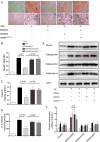
Figure 1. The anti-calcification effect of rhGHRH and GHRH-A (MR409) on SMCs from human aorta
A) SMCs from human aorta were cultured in osteogenic medium for 2-3 weeks and calcification were visualized by Alizarin Red staining. Both rhGHRH and MR409 inhibited calcium phosphate deposition. B) Quantification of Alizarin Red to represent the amount of calcium (*/#p<0.01, n=3). C) Total calcium was quantified (*/#p<0.01, n=3). Data were collected from three independent experiments. OM, osteogenic medium; rhGHRH, recombinant human growth hormone-releasing hormone; MR409, agonist of GHRH; MIA602, antagonist of GHRH. * was compared to “OM+rhGHRH” and # was compared to “OM+MR409”.
Figure 2
Figure 2. MR409 attenuates vascular calcification in OPG−/− mice
A) Micro-CT scans of thoracic aorta and 3D reconstruction. Upper panel, soft and calcific tissue were shown; Lower panel, only calcific area without soft tissue were presented; Red indicates soft tissue while calcification is indicated as white. B) Calcification (arrowhead) in aorta were visualized by Alizarin Red (upper panel) and Von Kossa (lower panel) stainings. C) Quantification of calcium deposition in A (*p<0.01). For OPG−/− mice group, 12 out of 17 mice in NC group and 11 out of 18 in MR409 treated group developed aorta calcification and quantified. D) Calcium and E) ALP activity in aorta were measured and were all significantly reduced by MR409 (*p<0.05). F) Representative Western blot analysis of Runx2, osteopontin, osteonectin and osteocalcin in aorta of mice. MR409 significantly impeded the upregulation of Runx2, osteonectin and osteocalcin expression in OPG−/− mice, but did not change the expression of osteopontin. G) Quantification of the proteins in F.
Figure 3
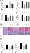
Figure 3. In vitro anti-calcification effect of MR409 on mSMCs
Mouse SMCs were cultured under OM with or without MR409 for 2-3 weeks. A) Calcific nodules were visualized by Alizarin Red (upper panel) and Von Kossa (lower panel) staining; B) Alizarin Red in A was quantified by measuring OD of the cell extraction (p<0.01); C) Total calcium and D) Alkaline phosphatase activity was measured (both p<0.001). E,F) Western blot analysis of Runx2, osteopontin, osteonectin and osteocalcin. MR409 significantly blocked the increase of Runx2, osteonectin and osteocalcin under OM (*p<0.01), and this effect can be attenuated by either the knock-down of GHRH-R by specific siRNA or the addition of GHRH antagonist, MIA602. However, osteopontin was not influenced by MR409. All data were collected from three independent experiments. AR, Alizarin Red staining; VK, Von Kossa staining. OM, osteogenic medium; MR409, agonist of GHRH; MIA602, antagonist of GHRH. * was compared to “OM+MR409” group.
Figure 4
Figure 4. ROS production and NADPH oxidase activity in mouse SMCs were reduced by MR409
A) ROS in SMCs were detected by DHE staining and observed by fluorescence microscope. The intensity of red fluorescence signal indicated the ROS content. B) The quantification of ROS content in A (*p<0.001, n=3, three independent experiments); C) NADPH oxidase activities in differently treated SMCs were measured by Luminescence Assay. Under OM conditions, ROS content and NADPH oxidase activity was significantly increased, and this was alleviated by MR409. The inhibition of ROS and NADPH oxidase activity by MR409 was reversed by treatments with the antagonist MIA602 or by siRNA against GHRH-R (*p<0.001, n=3, three independent experiments). NC, negative control; OM, osteogenic medium; MR409, agonist of GHRH; MIA602, antagonist of GHRH. * was compared to “OM+MR409” group.
Figure 5
Figure 5. PKA mediated de-activation of NADPH oxidase and ROS production
A) Cytosolic cAMP in mouse SMCs at specified culture conditions. cAMP was significantly elevated after MR409 treatment, and the effect was partially abolished by addition of MIA602 (*p<0.01, n=3); B) Measurement of NADPH oxidase activity in mSMCs. The enhanced activity of NADPH oxidase under OM condition was abolished by MR409 (*p<0.001, n=3). Inhibition of PKA by siRNA or inhibitor H89 blocked the effect of MR409 (§/&p<0.01, n=3); C) Quantification of ROS and D) Quantification of total calcium in mSMCs at specified culture conditions (*p<0.01, §/&p<0.001, n=3). E) Detection of calcification in mSMCs by Alizarin Red and Von Kossa staining. MR409 reduced calcium deposition under OM conditions, which was blocked by PKA inhibition. F) Measurement of NADPH oxidase activity in hSMCs. Inhibition of PKA by H89 in human SMCs also abolishes the MR409-mediated effect of NADPH oxidase de-activation (*p<0.0001, &p<0.001, n=3). G) Measurement of ROS in hSMCs. Blockage of ROS production by MR409 was attenuated when PKA was inhibited by H89 (*p<0.01, &p<0.05, n=3). All data were from three independent experiments. *,§, & were compared to the “OM+MR409” group.
Figure 6
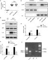
Figure 6. Inhibition of PKA confers dephosphorylation of NFκB (p65) in vitro
A) Western blot analysis of proteins in SMCs from WT mouse aorta. Under OM conditions, p-p65, IL-6 and Runx2 were increased, and were downregulated by MR409. The effects of MR409 were abolished by PKA knock-down. B,C,D) Quantification of p-p65,IL-6 and Runx2 expression in A (*p<0.01, n=3). E) mRNA level of RANKL of WT mice SMCs under OM conditions and MR409 did not influence OM mediated RANKL increase. F) Western blot analysis of proteins in SMCs from WT and OPG−/− mouse aorta. MR409 decreased the phosphorylation of p65 and the expression of IL-6 in the absence of OM and these effects were PKA dependent. G) Quantification of p-p65 and IL-6 expression in F (*p<0.01, n=3). All data were from three independent experiments. * was compared to the other groups.
Figure 7
Figure 7. MR409 promotes the dephosphorylation of NFκB in aorta of OPG−/− mouse
A) Serum RANKL in WT and OPG−/− mice treated with or without MR409. The OPG−/− mice possess higher serum level of RANKL, which was not altered by MR409 treatment. B) Western blot analysis of p-p65, p65, and IL-6 in aorta of WT and OPG−/− mice treated or not treated with MR409. Treatment of OPG−/− mice with MR409 reduced the phosphorylation of p65 and the expression of IL-6 in aorta. C) Quantification of p-p65 and IL-6 in B. D) Western blot of p-p65 in cytoplasm and nucleus. MR409 significantly reducd the amount of p-p65 both in cytoplasm and nucleus. E) Quantification of p-p65 D. F) CHIP assay showed the binding of p65 to the promoter region of Runx2.
Figure 8
Figure 8. Schematic diagram of the anti-calcification effect of GHRH-A
The binding of GHRH-A (MR409) to GHRH receptor activates the cAMP/PKA signaling pathway, resulting in inhibition of NADPH oxidase activity and dephosphorylation of NFκB (p65), which leads to the reduction of ROS and IL-6. Such effects of GHRH-A can be blocked by GHRH antagonist, MIA602, by siRNA of GHRH-R and PKA, and by chemical inhibitor H89. Reduction of ROS attenuates the expression of osteogenic genes such as transcription factor Runx2, osteonectin, osteocalcin and alkaline phosphatase. Finally, these changes lead to the inhibition of calcium phosphate deposition.

References

    1. Virmani R, Burke AP, Farb A, Kolodgie FD. Pathology of the vulnerable plaque. J Am Coll Cardiol. 2006;47:C13–18. - PubMed
    1. Abedin M, Tintut Y, Demer LL. Vascular calcification: Mechanisms and clinical ramifications. Arterioscler Thromb Vasc Biol. 2004;24:1161–1170. - PubMed
    1. Huang H, Virmani R, Younis H, Burke AP, Kamm RD, Lee RT. The impact of calcification on the biomechanical stability of atherosclerotic plaques. Circulation. 2001;103:1051–1056. - PubMed
    1. Vengrenyuk Y, Carlier S, Xanthos S, Cardoso L, Ganatos P, Virmani R, Einav S, Gilchrist L, Weinbaum S. A hypothesis for vulnerable plaque rupture due to stress-induced debonding around cellular microcalcifications in thin fibrous caps. Proc Natl Acad Sci U S A. 2006;103:14678–14683. - PMC - PubMed
    1. Aikawa E, Nahrendorf M, Figueiredo JL, Swirski FK, Shtatland T, Kohler RH, Jaffer FA, Aikawa M, Weissleder R. Osteogenesis associates with inflammation in early-stage atherosclerosis evaluated by molecular imaging in vivo. Circulation. 2007;116:2841–2850. - PubMed

Publication types

MeSH terms

Substances