. 2018 May 29;92(12):e00074-18.
doi: 10.1128/JVI.00074-18. Print 2018 Jun 15.

Large-Scale Complete-Genome Sequencing and Phylodynamic Analysis of Eastern Equine Encephalitis Virus Reveals Source-Sink Transmission Dynamics in the United States

Affiliations

Large-Scale Complete-Genome Sequencing and Phylodynamic Analysis of Eastern Equine Encephalitis Virus Reveals Source-Sink Transmission Dynamics in the United States

Yi Tan et al. J Virol. .

Abstract

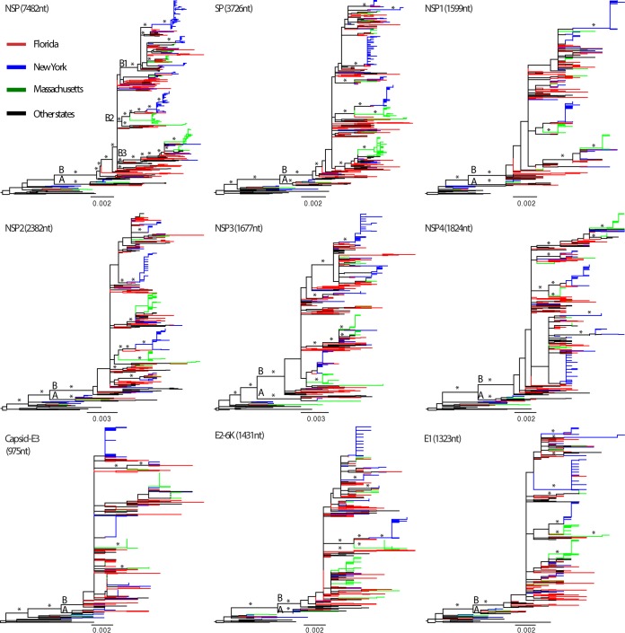
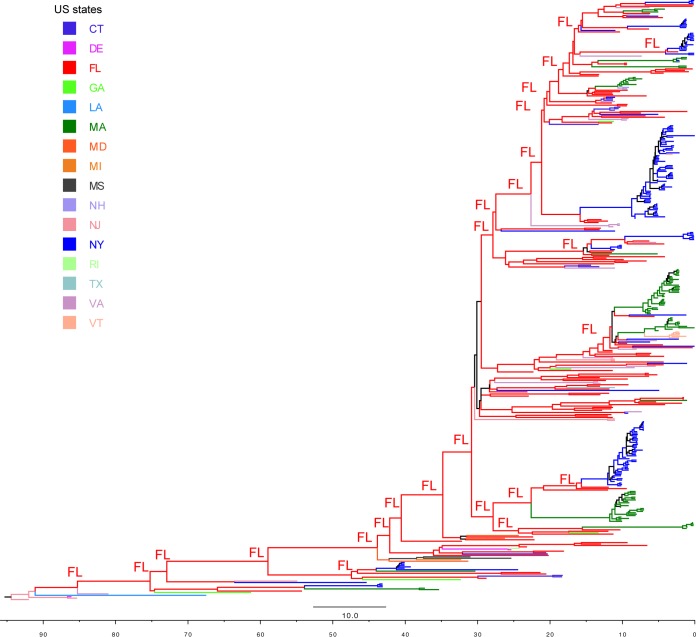
Eastern equine encephalitis virus (EEEV) has a high case-fatality rate in horses and humans, and Florida has been hypothesized to be the source of EEEV epidemics for the northeastern United States. To test this hypothesis, we sequenced complete genomes of 433 EEEV strains collected within the United States from 1934 to 2014. Phylogenetic analysis suggested EEEV evolves relatively slowly and that transmission is enzootic in Florida, characterized by higher genetic diversity and long-term local persistence. In contrast, EEEV strains in New York and Massachusetts were characterized by lower genetic diversity, multiple introductions, and shorter local persistence. Our phylogeographic analysis supported a source-sink model in which Florida is the major source of EEEV compared to the other localities sampled. In sum, this study revealed the complex epidemiological dynamics of EEEV in different geographic regions in the United States and provided general insights into the evolution and transmission of other avian mosquito-borne viruses in this region.IMPORTANCE Eastern equine encephalitis virus (EEEV) infections are severe in horses and humans on the east coast of the United States with a >90% mortality rate in horses, an ∼33% mortality rate in humans, and significant brain damage in most human survivors. However, little is known about the evolutionary characteristics of EEEV due to the lack of genome sequences. By generating large collection of publicly available complete genome sequences, this study comprehensively determined the evolution of the virus, described the epidemiological dynamics of EEEV in different states in the United States, and identified Florida as one of the major sources. These results may have important implications for the control and prevention of other mosquito-borne viruses in the Americas.

Keywords: EEEV; NGS; evolution; next-generation sequencing; phylodynamics; phylogeography; source-sink dynamics.


Figures

FIG 1
FIG 1
Evolution of EEEV genomes. (A) Entropy analyses of the genomic nucleotide sequences (blue bars) and translated amino acid sequences (red bars) of EEEV isolates (n = 437) from 1934 to 2014. Estimates of dN/dS (green line) of the sliding windows (window size = 30 codons) across the genome. The x axis is the genomic position, while the left y axis is the entropy level, and the right y axis is dN/dS. (B) Evolutionary rates (nucleotide substitutions/site/year) of the complete genome and each individual gene. They were estimated using a Bayesian protocol and displayed in boxplot where the whiskers extend to the 95% highest posterior density (HPD) interval, and the boxes indicate the 25 to 75% interquartile range of the posterior distribution of rate estimates, thus describing its central tendency. (C) Root-to-tip regression analysis for complete genome sequences. Dash lines are the 95% prediction bands of the regression. Frequencies of sequences sampled in different states and time are summarized in the stacked bar chart, with the y axis shown on the right.
FIG 2
FIG 2
Threading of EEEV structural polyprotein through an endoplasmic reticulum membrane. Structure proteins are colored separately: Capsid in yellow, E3 in green, E2 in blue, 6K in red, and E1 in magenta. The positive selection pressure site, residue 45 in 6K, is indicated by a black arrow.
FIG 3
FIG 3
Time-scaled phylogeny of EEEV. A maximum clade credibility (MCC) tree of complete genome sequences of EEEV is shown. Major nodes with posterior probabilities over 0.9 are marked by asterisks. Sequences from Florida, New York, Massachusetts, and other places are colored and described in the legend key. Clades A and B and subclades B1, B2, and B3, as well as the 19 monophyletic groups further defined for Florida, New York, and Massachusetts viruses, are shown. The times of the most recent common ancestors (tMRCAs) of all EEEVs and the monophyletic groups are provided after the colons. Mutations at residue 45 (Ala→Vla) in the 6K protein are mapped on the branches of the tree using yellow circles. The inset diagram on the left shows the time spans of these monophyletic groups, including the range of the sampling time (rectangular boxes) and the mean of the tMRCAs (vertical arrow bars).
FIG 4
FIG 4
ML phylogenetic tree of complete genome sequences of EEEV. Well-supported nodes by bootstrap values over 70% and posterior probabilities > 0.9 from BMCMC analyses are marked by asterisks. Clades (A and B) and subclades (B1, B2, and B3) are described in the trees. Sequences sampled from Florida, New York, and Massachusetts are shown in red, blue, and green, respectively, and described in the key. The monophyletic groups of Florida, New York, and Massachusetts sequences are indicated in the trees. Phylogenetic trees were rooted by the oldest EEEV strain, Ten Broeck, collected in 1933 in Virginia. Scale bars represent the number of nucleotide substitutions per site.
FIG 5
FIG 5
Phylogenetic trees of individual genes of EEEV. ML phylogenetic trees inferred for different regions of the EEEV genome are indicated in black. Well-supported nodes by bootstrap values over 70% are marked by asterisks. Sequences from Florida, New York, Massachusetts, and other places are colored and described in the key. Phylogenetic trees were rooted using the oldest EEEV strain, Ten Broeck, collected in 1933, and scale bars represent the number of nucleotide substitutions per site.
FIG 6
FIG 6
Inference of the ancestral states in the MCC tree of EEEV. Sequences from different states are colored and described in the key. The ancestor geographic states with state probabilities over 0.9 at the backbone of the EEEV phylogeny are labeled in the tree.
FIG 7
FIG 7
Map of the United States showing the EEEV cases and the virus movement inferred from the Bayesian phylogeographic analysis. Red circles in each state represent the EEEV isolates collected during 1931 to 2014 (circle size is proportional to the number of sequences analyzed). The numbers of human cases are indicated by the yellow shade in each state. Green arrow lines indicate the significant virus movement between states as reported by Bayes factor > 20 in the phylogeographic analysis using BEAST program. The line width is proportional to the transition rate between the states, as shown in Table 2.

References

    1. Powers AM, Brault AC, Shirako Y, Strauss EG, Kang W, Strauss JH, Weaver SC. 2001. Evolutionary relationships and systematics of the alphaviruses. J Virol 75:10118–10131. doi: 10.1128/JVI.75.21.10118-10131.2001. - DOI - PMC - PubMed
    1. Weaver SC, Winegar R, Manger ID, Forrester NL. 2012. Alphaviruses: population genetics and determinants of emergence. Antiviral Res 94:242–257. doi: 10.1016/j.antiviral.2012.04.002. - DOI - PMC - PubMed
    1. Feemster RF. 1938. Outbreak of encephalitis in man due to the eastern virus of equine encephalomyelitis. Am J Public Health Nations Health 28:1403–1410. doi: 10.2105/AJPH.28.12.1403. - DOI - PMC - PubMed
    1. Fothergill LD, Dingle JH, Fellow JJ. 1938. A fatal disease of pigeons caused by the virus of the eastern variety of equine encephalomyelitis. Science 88:549–550. doi: 10.1126/science.88.2293.549-a. - DOI - PubMed
    1. Webster LT, Wright FH. 1938. Recovery of eastern equine encephalomyelitis virus from brain tissue of human cases of encephalitis in Massachusetts. Science 88:305–306. doi: 10.1126/science.88.2283.305. - DOI - PubMed

Publication types

LinkOut - more resources