. 2018 Apr 5;3(7):e98575.
doi: 10.1172/jci.insight.98575.

Comprehensive immunoproteogenomic analyses of malignant pleural mesothelioma

Affiliations

Comprehensive immunoproteogenomic analyses of malignant pleural mesothelioma

Hyun-Sung Lee et al. JCI Insight. .

Abstract

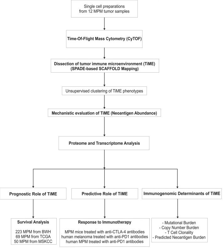
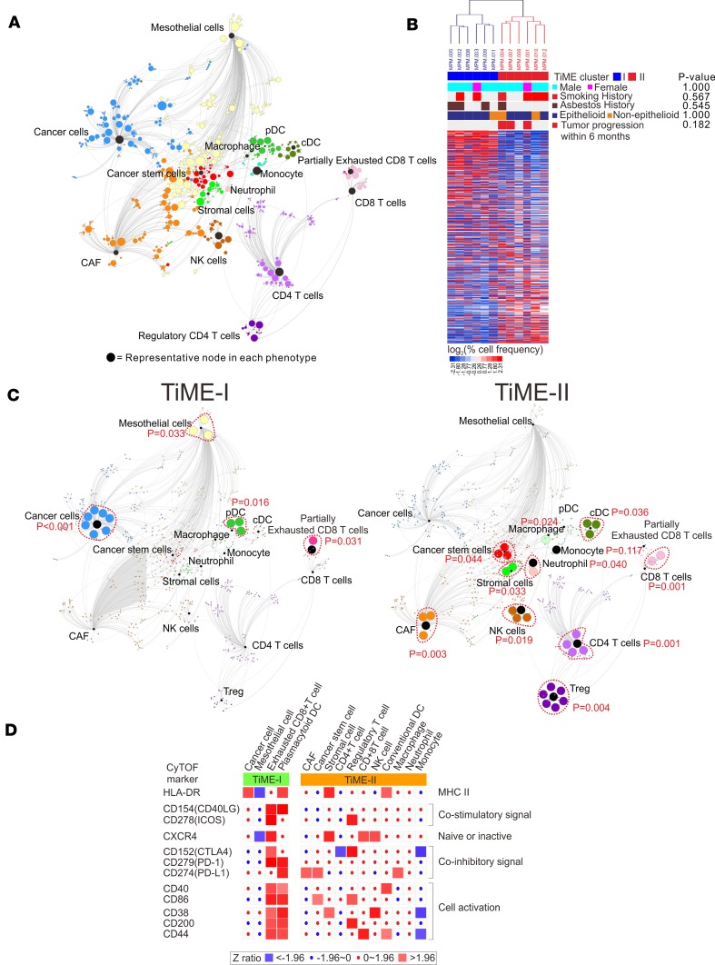
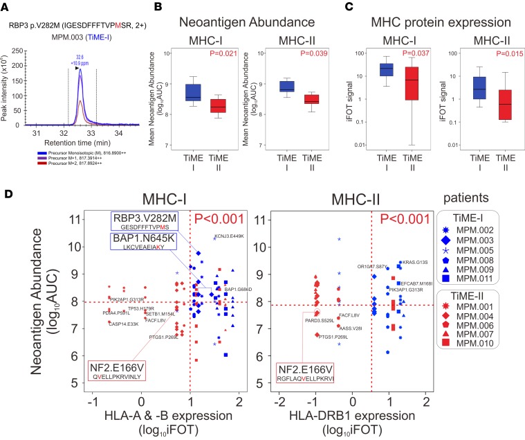
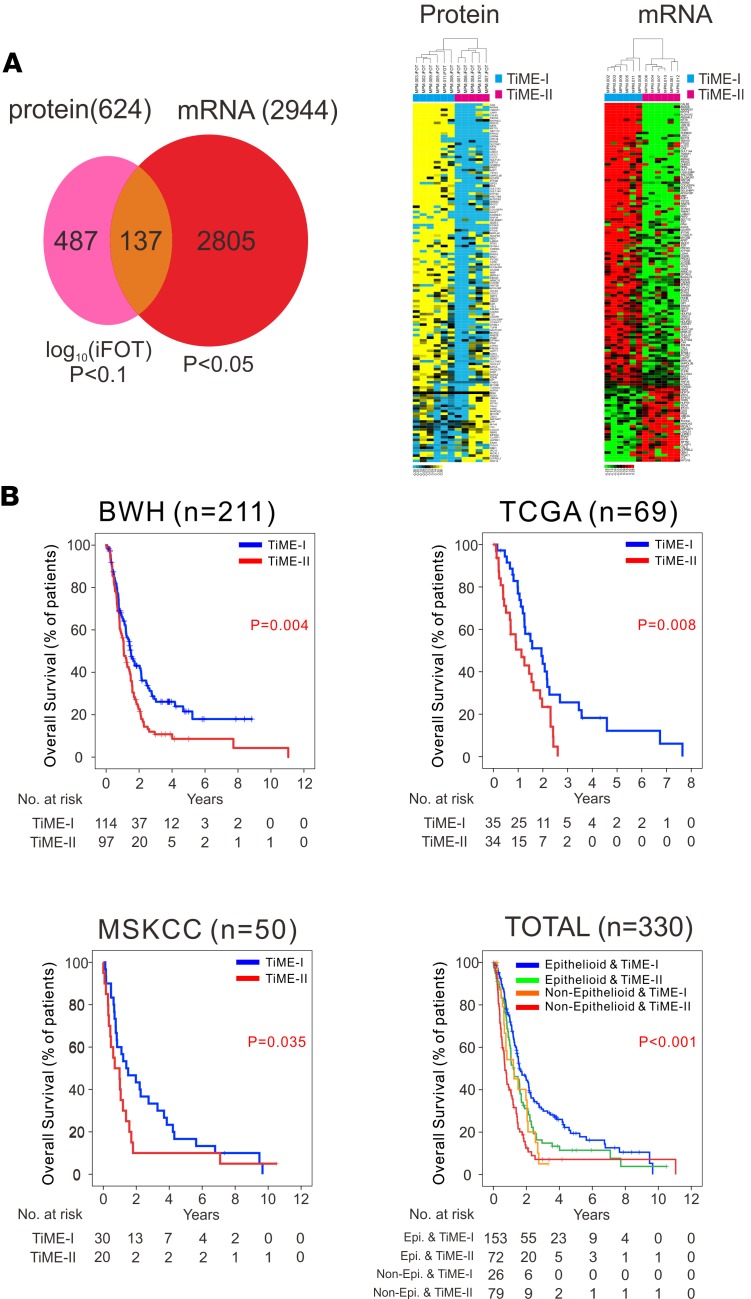
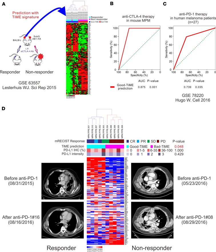
We generated a comprehensive atlas of the immunologic cellular networks within human malignant pleural mesothelioma (MPM) using mass cytometry. Data-driven analyses of these high-resolution single-cell data identified 2 distinct immunologic subtypes of MPM with vastly different cellular composition, activation states, and immunologic function; mass spectrometry demonstrated differential abundance of MHC-I and -II neopeptides directly identified between these subtypes. The clinical relevance of this immunologic subtyping was investigated with a discriminatory molecular signature derived through comparison of the proteomes and transcriptomes of these 2 immunologic MPM subtypes. This molecular signature, representative of a favorable intratumoral cell network, was independently associated with improved survival in MPM and predicted response to immune checkpoint inhibitors in patients with MPM and melanoma. These data additionally suggest a potentially novel mechanism of response to checkpoint blockade: requirement for high measured abundance of neopeptides in the presence of high expression of MHC proteins specific for these neopeptides.

Keywords: Cancer immunotherapy; Expression profiling; Immunology; Oncology; Proteomics.


Conflict of interest statement

Conflict of interest: The authors have declared that no conflict of interest exists.

Figures

Figure 1
Figure 1. Schematic illustration of study design.
BWH, Brigham and Women’s Hospital; CTLA-4, cytotoxic t-lymphocyte associated protein 4; CyTOF, time-of-flight mass cytometry; MPM, malignant pleural mesothelioma; MSKCC, Memorial Sloan Kettering Cancer Center; PD-1, programmed cell death 1; SCAFFOLD, single-cell analysis by fixed force– and landmark-directed; SPADE, spanning-tree progression analysis of density-normalized events; TCGA, The Cancer Genome Atlas; and TiME, tumor immune microenvironment.
Figure 2
Figure 2. SCAFFOLD maps of tumor immune microenvironment (TiME) in MPM.
(A) A SCAFFOLD map of TiME in 12 human MPM tumors. CyTOF was performed on 12 MPM tumors utilizing a panel of 35 metal-conjugated antibodies. Pooled data from these 12 patients was used to generate a SCAFFOLD reference map of MPM’s intratumoral immune system. This approach provides a data-driven representation of cellular networks, while also denoting the location of landmark immune cell populations defined using prior knowledge of the immune system. For example, landmark nodes are visualized as black nodes and represent 15 manually defined major cellular phenotypes. The same cells are subjected to unsupervised clustering to provide an objective view of cell composition and organization, and 742 cellular subpopulations were identified and represented by the colored nodes. In these maps, node size represents the relative number of cells in that grouping, and line length indicates similarity between cells. In other words, 2 groups of cells are connected by a short line if the proteins they express are relatively similar, and a longer line if they are relatively disparate. (B) Two distinct subsets of MPM patients were identified by unsupervised clustering of pooled CyTOF data from 12 MPM tumors: 6 tumors of the TiME-I subset and 6 tumors of the TiME-II subset. (C) The SCAFFOLD maps of TiME-I and TiME-II subsets. SCAFFOLD maps were generated from pooled data from the 6 patients in each of the TiME-I and -II immunologic subsets of MPM, and cellular subpopulations were statistically compared between each subset. The internodal differences in the same phenotypes were analyzed with 2-tailed paired t test according to the corresponding nodes. (D) Differential activation states of the immune cell populations between TiME-I and TiME-II MPM tumors. Immune stimulatory or inhibitory markers were significantly altered between 2 TiME subsets. Z ratios were calculated by taking the difference between the averages of the observed marker Z scores and dividing by the SD of all the differences for that particular comparison. A Z ratio of ±1.96 was inferred as significant (P < 0.05). CAF, cancer-associated fibroblast; cDC, conventional DCs; CyTOF, time-of-flight mass cytometry; MPM, malignant pleural mesothelioma; pDC, plasmacytoid DCs; SCAFFOLD, single-cell analysis by fixed force– and landmark-directed; TiME, tumor immune microenvironment; and Treg, CD4+ Tregs.
Figure 3
Figure 3. Neoantigen abundance and corresponding MHC molecules between 2 distinct TiME subsets.
(A) Direct identification of neoantigen abundance of IGESDFFFTVPMSR of RBP3V282M by mass spectrometry. Triple-redundant peaks of monoisotopic 12C, 13C (M+1), and 14C (M+2) support that the identified peaks for the peptides are accurately made. (B) Mean neoantigen abundance of directly identified neopeptides for MHC-I and MHC-II was determined by mass spectrometry on 11 MPM tumors (n = 6 TIME-I and n = 5 TiME-II). The 2-tailed Student’s t tests were used to compare the data. (C) MHC-I and MHC-II protein expression was determined by mass spectrometry on 11 MPM tumors. The 2-tailed Student’s t tests were used to compare the data. (D) Two-dimensional plots between abundance of neopeptides with high affinity to HLA-A, HLA-B, and HLA-DRB1 and the expression of the specific corresponding MHC molecules, on 11 MPM tumors. The 2-tailed χ2 tests were used to compare the data. BAP1, BRCA1 associated protein 1; iFOT, fraction of total intensity based absolute quantification; MHC, major histocompatibility complex; MPM, malignant pleural mesothelioma; NF2, neurofibromin 2; RBP3, retinol binding protein 3; and TiME, tumor immune microenvironment.
Figure 4
Figure 4. Prognostic significance of the TiME signature in MPM.
(A) Development of a molecular signature that discriminates TiME-I and TiME-II MPM tumors through protein profiling by mass spectrometry and mRNA transcriptome analysis using 137 differential proteins, also differentially expressed in mRNA. (B) Kaplan-Meier curves of overall survival in the BWH cohort (n = 211), the TCGA MPM cohort (n = 69), the MSKCC cohort (n = 50), and a combined dataset (n = 330). Survival curves were generated with the Kaplan-Meier’s method, and intergroup comparisons were performed with the log-rank test. BWH, Brigham and Women’s Hospital; MPM, malignant pleural mesothelioma; MSKCC, Memorial Sloan Kettering Cancer Center; TCGA, The Cancer Genome Atlas; and TiME, tumor immune microenvironment.
Figure 5
Figure 5. The TiME signature to predict response to immune checkpoint inhibitors.
(A) Predictive role of the TiME signature in a mouse MPM model treated with anti–CTLA-4 antibodies. (B) AUC analysis of TiME signature in a mouse MPM model treated with anti–CTLA-4 antibodies. (C) Predictive role of the TiME signature in a cohort of patients with advanced melanoma treated with PD-1 blockade (n = 27). (D) Predictive role of the TiME signature in 10 unresectable human MPM patients treated with anti–PD-1 therapy. The 2-tailed Fisher’s exact tests were used to compare the data. CR, complete response; CTLA-4, cytotoxic T-lymphocyte associated protein 4; MPM, malignant pleural mesothelioma; mRECIST, modified response evaluation criteria in solid tumors; PD, progressive disease; PD-L1, programmed cell death 1 ligand 1; PD-1, programmed cell death 1; PR, partial response; SD, stable disease; and TiME, tumor immune microenvironment.

References

    1. Merritt N, Blewett CJ, Miller JD, Bennett WF, Young JE, Urschel JD. Survival after conservative (palliative) management of pleural malignant mesothelioma. J Surg Oncol. 2001;78(3):171–174. doi: 10.1002/jso.1143. - DOI - PubMed
    1. Zalcman G, et al. Bevacizumab for newly diagnosed pleural mesothelioma in the Mesothelioma Avastin Cisplatin Pemetrexed Study (MAPS): a randomised, controlled, open-label, phase 3 trial. Lancet. 2016;387(10026):1405–1414. doi: 10.1016/S0140-6736(15)01238-6. - DOI - PubMed
    1. Sugarbaker DJ, Richards WG, Bueno R. Extrapleural pneumonectomy in the treatment of epithelioid malignant pleural mesothelioma: novel prognostic implications of combined N1 and N2 nodal involvement based on experience in 529 patients. Ann Surg. 2014;260(4):577–582. doi: 10.1097/SLA.0000000000000903. - DOI - PMC - PubMed
    1. Quispel-Janssen J, et al. A phase II study of nivolumab in malignant pleural mesothelioma (NivoMes): with translational research (TR) biopsies. J Thorac Oncol. 2017;12:S292–S293.
    1. Kindler H, et al. Phase II trial of pembrolizumab in patients with malignant mesothelioma (MM): interim analysis. J Thorac Oncol. 2017;12:S293–S294.

Publication types

MeSH terms

Substances