. 2018 Apr 4;8(1):5594.
doi: 10.1038/s41598-018-23908-z.

Respiration and the watershed of spinal CSF flow in humans

Affiliations

Respiration and the watershed of spinal CSF flow in humans

Steffi Dreha-Kulaczewski et al. Sci Rep. .

Abstract

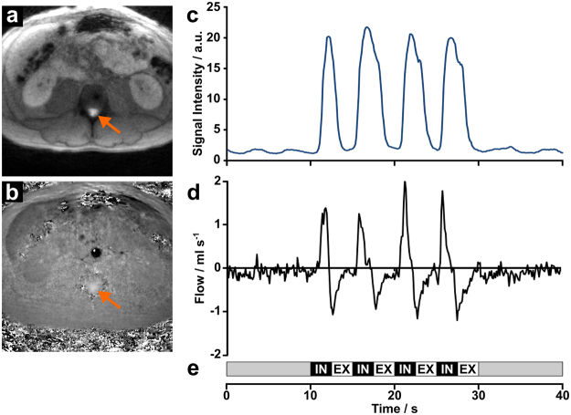
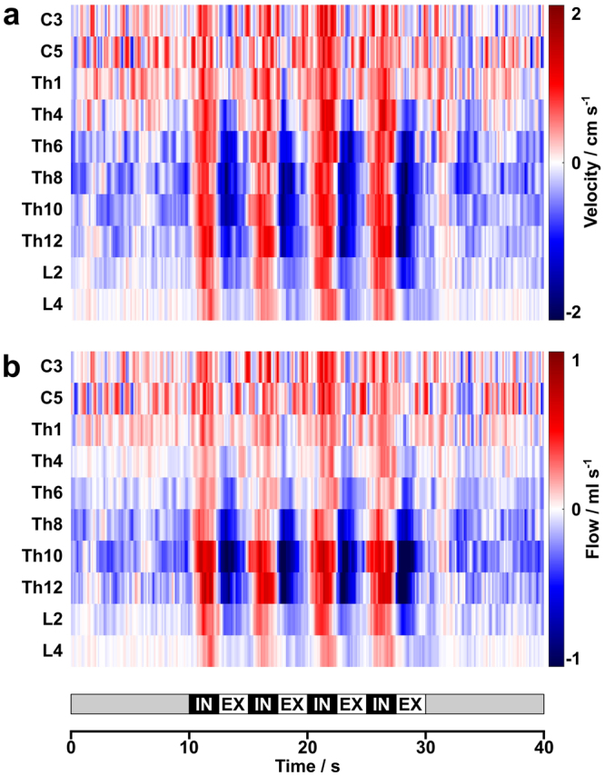
The dynamics of human CSF in brain and upper spinal canal are regulated by inspiration and connected to the venous system through associated pressure changes. Upward CSF flow into the head during inspiration counterbalances venous flow out of the brain. Here, we investigated CSF motion along the spinal canal by real-time phase-contrast flow MRI at high spatial and temporal resolution. Results reveal a watershed of spinal CSF dynamics which divides flow behavior at about the level of the heart. While forced inspiration prompts upward surge of CSF flow volumes in the entire spinal canal, ensuing expiration leads to pronounced downward CSF flow, but only in the lower canal. The resulting pattern of net flow volumes during forced respiration yields upward CSF motion in the upper and downward flow in the lower spinal canal. These observations most likely reflect closely coupled CSF and venous systems as both large caval veins and their anastomosing vertebral plexus react to respiration-induced pressure changes.


Conflict of interest statement

The authors declare no competing interests.

Figures

Figure 1
Figure 1
Real-time phase-contrast flow MRI of the spinal canal. (a) Selected anatomic image at lumbar level L4 depicting CSF through-plane flow (arrow) and (b) corresponding velocity map with bright signals (arrow) indicating CSF flow in upward direction. (c) Signal intensity in the abdominal wall from anatomic images indicating the subjects breathing performance and (d) corresponding CSF flow (ml s−1) in response to (e) a visually presented breathing protocol comprising 4 cycles of 2.5 s forced inspiration (IN) and 2.5 s expiration (EX).
Figure 2
Figure 2
CSF dynamics along the spinal canal. (a) Mean color-coded flow velocities and (b) flow rates averaged across subjects as a function of spinal level. CSF flow increases in upward direction (red) with every forced inspiration at all spinal levels. During subsequent expiration CSF dynamics follow a bidirectional pattern with very low flow in the upper spinal canal and a pronounced downward movement (blue) in the lower spinal canal. White color represents zero flow.
Figure 3
Figure 3
Watershed of spinal CSF flow during forced breathing. (a) Mean CSF volumes averaged across subjects from all 4 cycles of forced inspiration (IN = red) and expiration (EX = blue) as a function of spinal level. While inspiratory CSF volumes represent upward flow in the entire spinal canal, expiration leads to a downward directionality in the lower spinal canal. (b) Corresponding net CSF flow volumes point upwards at cervical and high thoracic levels and downwards between mid and lower thoracic regions. Error bars represent standard deviations.

References

    1. Dichiro G. Movement of the Cerebrospinal Fluid in Human Beings. Nature. 1964;204:290–291. doi: 10.1038/204290a0. - DOI - PubMed
    1. Di Chiro G. Observations on the circulation of the cerebrospinal fluid. Acta Radiol Diagn (Stockh) 1966;5:988–1002. doi: 10.1177/02841851660050P242. - DOI - PubMed
    1. Chiro GD, Hammock MK, Bleyer WA. Spinal descent of cerebrospinal fluid in man. Neurology. 1976;26:1–8. doi: 10.1212/WNL.26.1.1. - DOI - PubMed
    1. Greitz D, Hannerz J. A proposed model of cerebrospinal fluid circulation: observations with radionuclide cisternography. AJNR Am J Neuroradiol. 1996;17:431–438. - PMC - PubMed
    1. Greitz D, Franck A, Nordell B. On the pulsatile nature of intracranial and spinal CSF-circulation demonstrated by MR imaging. Acta Radiol. 1993;34:321–328. doi: 10.1177/028418519303400403. - DOI - PubMed