

Results of the radiation dose of head, body and tail of hippocampus in nasopharyngeal carcinoma patients treated with intensity modulated radiotherapy
- PMID: 29618828
- PMCID: PMC5884782
- DOI: 10.1038/s41598-018-23127-6
Results of the radiation dose of head, body and tail of hippocampus in nasopharyngeal carcinoma patients treated with intensity modulated radiotherapy
Abstract
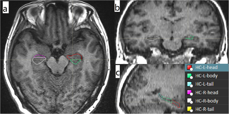
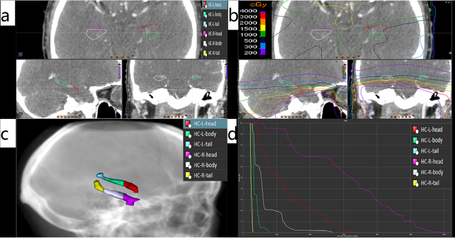
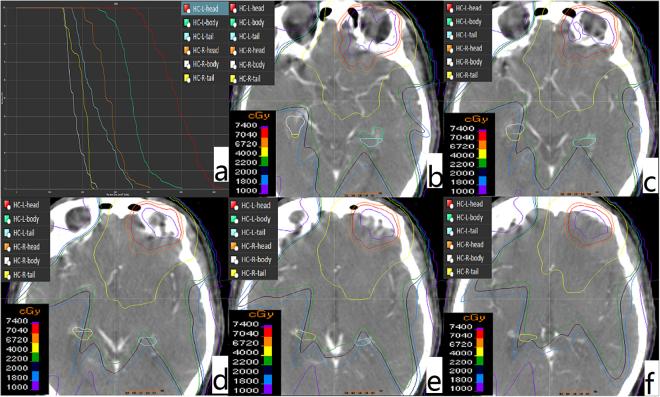
This study is to analyze the radiation dose of head, body and tail of hippocampus (HC) of nasopharyngeal carcinoma (NPC) patients treated with intensity modulated radiotherapy (IMRT). Evaluate cognitive function of patients with Wechsler adult intelligence scale-Chinese revised (WAIS-CR). HC were segmented into HC head (HH), HC body (HB) and HC tail (HT) and the indexes were then analyzed. WAIS-CR was tested before and 3months after radiotherapy. The mean radiation dose of left and right HC was (1147 ± 976)cGy, (1011 ± 602)cGy respectively. The radiation dose and the volume exposed in different dose of HH, HB and HT decreased in turn. For 5 patients, before and after radiotherapy, the regular-order score was 8.60 ± 1.34, 8.0 ± 1.00 (P = 0.43), while the reverse-order score was 5.80 ± 0.84, 5.20 ± 0.84 (P = 0.07). The radiation dose of HH, HB and HT was different, and the radiation dose of HH was the highest, which should be emphasized especially.
Conflict of interest statement
The authors declare no competing interests.
Figures
References
-
- Gondi V, et al. Preservation of memory with conformal avoidance of the hippocampal neural stem-cell compartment during whole-brain radiotherapy for brain metastases (RTOG 0933): a phase II multi-institutional trial. J Clin Oncol. 2014;32:3810–3816. doi: 10.1200/JCO.2014.57.2909. - DOI - PMC - PubMed
Publication types
MeSH terms
Substances
LinkOut - more resources
Full Text Sources
Other Literature Sources
