

Corticotropin-Releasing Factor Receptors Modulate Oxytocin Release in the Dorsolateral Bed Nucleus of the Stria Terminalis (BNST) in Male Rats
- PMID: 29618970
- PMCID: PMC5871712
- DOI: 10.3389/fnins.2018.00183
Corticotropin-Releasing Factor Receptors Modulate Oxytocin Release in the Dorsolateral Bed Nucleus of the Stria Terminalis (BNST) in Male Rats
Abstract
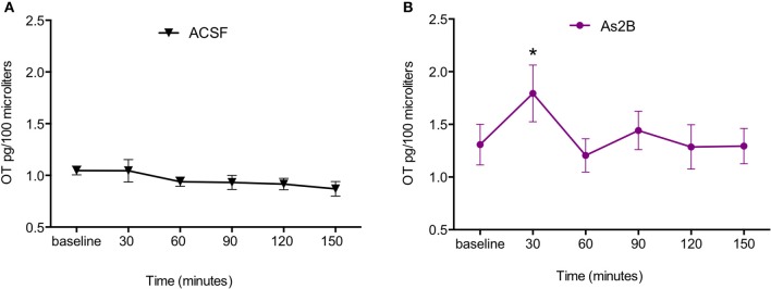
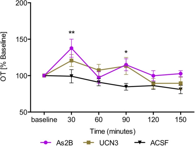
The neuropeptide oxytocin (OT) plays an important role in the regulation of social and anxiety-like behavior. Our previous studies have shown that OT neurons send projections from the hypothalamus to the dorsolateral bed nucleus of the stria terminalis (BNSTdl), a forebrain region critically involved in the modulation of anxiety-like behavior. Importantly, these OT terminals in the BNSTdl express presynaptic corticotropin releasing factor (CRF) receptor type 2 (CRFR2). This suggests that CRFR2 might be involved in the modulation of OT release. To test this hypothesis, we measured OT content in microdialysates collected from the BNSTdl of freely-moving male Sprague-Dawley rats following the administration of a selective CRFR2 agonist (Urocortin 3) or antagonist (Astressin 2B, As2B). To determine if type 1 CRF receptors (CRFR1) are also involved, we used selective CRFR1 antagonist (NBI35965) as well as CRF, a putative ligand of both CRFR1 and CRFR2. All compounds were delivered directly into the BNSTdl via reverse dialysis. OT content in the microdialysates was measured with highly sensitive and selective radioimmunoassay. Blocking CRFR2 with As2B caused an increase in OT content in BNSTdl microdialysates, whereas CRFR2 activation by Urocortin 3 did not have an effect. The As2B-induced increase in OT release was blocked by application of the CRFR1 antagonist demonstrating that the effect was dependent on CRFR1 transmission. Interestingly, CRF alone caused a delayed increase in OT content in BNSTdl microdialysates, which was dependent on CRF2 but not CRF1 receptors. Our results suggest that members of the CRF peptide family modulate OT release in the BNSTdl via a fine-tuned mechanism that involves both CRFR1 and CRFR2. Further exploration of mechanisms by which endogenous OT system is modulated by CRF peptide family is needed to better understand the role of these neuropeptides in the regulation of anxiety and the stress response.
Keywords: BNST; CRF; bed nucleus of the stria terminalis; microdialysis; oxytocin; release; urocortin.
Figures


Similar articles
-
Neuroanatomical evidence for reciprocal regulation of the corticotrophin-releasing factor and oxytocin systems in the hypothalamus and the bed nucleus of the stria terminalis of the rat: Implications for balancing stress and affect.Psychoneuroendocrinology. 2011 Oct;36(9):1312-26. doi: 10.1016/j.psyneuen.2011.03.003. Epub 2011 Apr 9. Psychoneuroendocrinology. 2011. PMID: 21481539 Free PMC article.
-
Oxytocin receptors in the dorsolateral bed nucleus of the stria terminalis (BNST) bias fear learning toward temporally predictable cued fear.Transl Psychiatry. 2019 Apr 18;9(1):140. doi: 10.1038/s41398-019-0474-x. Transl Psychiatry. 2019. PMID: 31000694 Free PMC article.
-
Oxytocin excites BNST interneurons and inhibits BNST output neurons to the central amygdala.Neuropharmacology. 2021 Jul 1;192:108601. doi: 10.1016/j.neuropharm.2021.108601. Epub 2021 May 7. Neuropharmacology. 2021. PMID: 33971215 Free PMC article.
-
Oxytocin facilitates adaptive fear and attenuates anxiety responses in animal models and human studies-potential interaction with the corticotropin-releasing factor (CRF) system in the bed nucleus of the stria terminalis (BNST).Cell Tissue Res. 2019 Jan;375(1):143-172. doi: 10.1007/s00441-018-2889-8. Epub 2018 Jul 28. Cell Tissue Res. 2019. PMID: 30054732 Free PMC article. Review.
-
Actions of CRF and its analogs.Curr Med Chem. 1999 Nov;6(11):1035-53. Curr Med Chem. 1999. PMID: 10519912 Review.
Cited by
-
Neuronal diversity of the amygdala and the bed nucleus of the stria terminalis.Handb Behav Neurosci. 2020;26:63-100. doi: 10.1016/b978-0-12-815134-1.00003-9. Epub 2020 Mar 31. Handb Behav Neurosci. 2020. PMID: 32792868 Free PMC article. No abstract available.
-
Corticotropin releasing factor and norepinephrine related circuitry changes in the bed nucleus of the stria terminalis in stress and alcohol and substance use disorders.Neuropharmacology. 2021 Dec 15;201:108814. doi: 10.1016/j.neuropharm.2021.108814. Epub 2021 Oct 6. Neuropharmacology. 2021. PMID: 34624301 Free PMC article. Review.
-
The modulation of emotional and social behaviors by oxytocin signaling in limbic network.Front Mol Neurosci. 2022 Nov 17;15:1002846. doi: 10.3389/fnmol.2022.1002846. eCollection 2022. Front Mol Neurosci. 2022. PMID: 36466805 Free PMC article. Review.
-
Hypoxia activates a neuropeptidergic pathway from the paraventricular nucleus of the hypothalamus to the nucleus tractus solitarii.Am J Physiol Regul Integr Comp Physiol. 2018 Dec 1;315(6):R1167-R1182. doi: 10.1152/ajpregu.00244.2018. Epub 2018 Sep 19. Am J Physiol Regul Integr Comp Physiol. 2018. PMID: 30230933 Free PMC article.
-
Extended amygdala corticotropin-releasing hormone neurons regulate sexually dimorphic changes in pair bond formation following social defeat in prairie voles (Microtus ochrogaster).Neuropsychopharmacology. 2025 May;50(6):965-975. doi: 10.1038/s41386-025-02067-6. Epub 2025 Feb 12. Neuropsychopharmacology. 2025. PMID: 39939823
References
-
- Bosch O. J., Dabrowska J., Modi M. E., Johnson Z. V., Keebaugh A. C., Barrett C. E., et al. . (2016). Oxytocin in the nucleus accumbens shell reverses CRFR2-evoked passive stress-coping after partner loss in monogamous male prairie voles. Psychoneuroendocrinology 64, 66–78. 10.1016/j.psyneuen.2015.11.011 - DOI - PMC - PubMed
Grants and funding
LinkOut - more resources
Full Text Sources
Other Literature Sources