Review
. 2018 Mar 21:9:257.
doi: 10.3389/fphys.2018.00257. eCollection 2018.

The Complex Role of Store Operated Calcium Entry Pathways and Related Proteins in the Function of Cardiac, Skeletal and Vascular Smooth Muscle Cells

Affiliations
Review

The Complex Role of Store Operated Calcium Entry Pathways and Related Proteins in the Function of Cardiac, Skeletal and Vascular Smooth Muscle Cells

Javier Avila-Medina et al. Front Physiol. .

Abstract

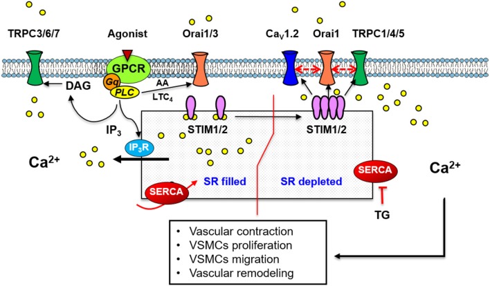
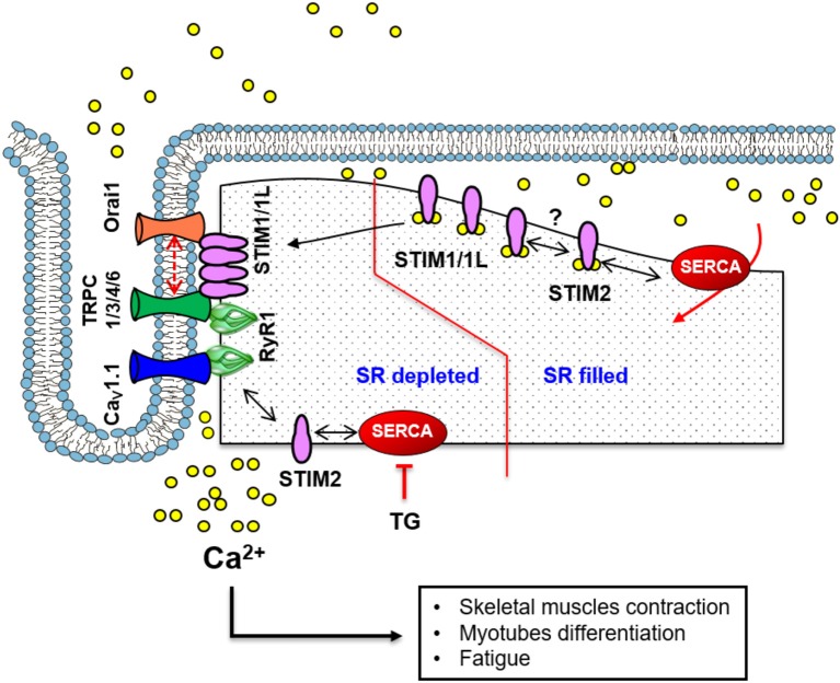
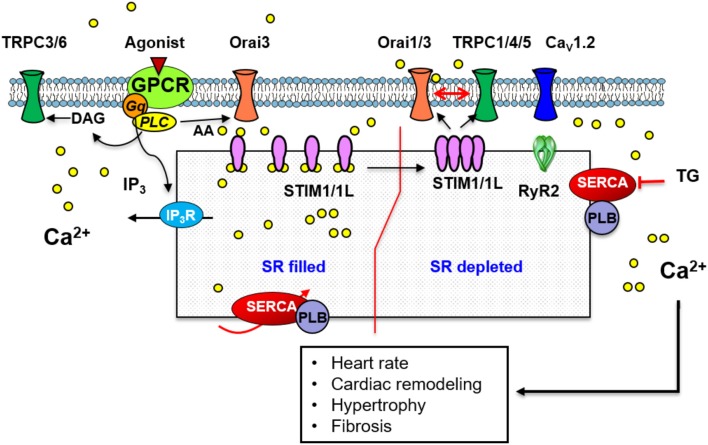
Cardiac, skeletal, and smooth muscle cells shared the common feature of contraction in response to different stimuli. Agonist-induced muscle's contraction is triggered by a cytosolic free Ca2+ concentration increase due to a rapid Ca2+ release from intracellular stores and a transmembrane Ca2+ influx, mainly through L-type Ca2+ channels. Compelling evidences have demonstrated that Ca2+ might also enter through other cationic channels such as Store-Operated Ca2+ Channels (SOCCs), involved in several physiological functions and pathological conditions. The opening of SOCCs is regulated by the filling state of the intracellular Ca2+ store, the sarcoplasmic reticulum, which communicates to the plasma membrane channels through the Stromal Interaction Molecule 1/2 (STIM1/2) protein. In muscle cells, SOCCs can be mainly non-selective cation channels formed by Orai1 and other members of the Transient Receptor Potential-Canonical (TRPC) channels family, as well as highly selective Ca2+ Release-Activated Ca2+ (CRAC) channels, formed exclusively by subunits of Orai proteins likely organized in macromolecular complexes. This review summarizes the current knowledge of the complex role of Store Operated Calcium Entry (SOCE) pathways and related proteins in the function of cardiac, skeletal, and vascular smooth muscle cells.

Keywords: Ca2+; Orai; STIM; TRPC; cardiomyocyte; skeletal muscle; vascular smooth muscle.


Figures

Figure 1
Figure 1
A scheme illustrating the standard mechanism and molecular components of SOCE in vascular smooth muscle cells. Store depletion by thapsigargin or vasoactive agonists binding to GPCR, induces Ca2+ store depletion, STIM1 organization in puncta and translocation to the plasma membrane, activation of Ca2+ entry through Orai1 and/or TRPC-dependent SOCC. A mechanism involving Orai1, TRPC1 and the voltage-sensitive Cav1.2, has also been implicated in Ca2+ influx induced by SOCE. Agonists also stimulate store independent Ca2+ entry through Orai1/3 activated by AA or LTC4; or through TRPC3/6/7 activated by DAG.
Figure 2
Figure 2
A scheme illustrating the standard mechanism and molecular components of SOCE in skeletal muscle. At the triad junction, the voltage-sensitive CaV1.1 and RyR1 interacts physically. STIM1 and STIM1L are in close proximity or in complex with Orai1 even before store depletion. This pre-localization of STIM1/1L with Orai1 likely facilitates the fast-activation kinetics of SOCE. A similar mechanism involving TRPC channels and STIM1 has also been implicated in Ca2+ influx mechanisms. STIM1 interacts with STIM2, which regulates SERCA1-mediated filling of the SR Ca2+ store and sustains resting [Ca2+] in the cytosol.
Figure 3
Figure 3
A scheme illustrating the standard mechanism and molecular components of SOCE in cardiac muscle. Store depletion involves STIM1/1L interaction with Orai1 which likely recruits Orai3. STIM1/1L colocalizes with key regulators of SOCE such as Orai1, SERCA, phospholamban (PLB), and RyR2. A similar mechanism involving TRPC1/4/5 channels and STIM1 has also been implicated in SOCE. Store independent Ca2+ entry through Orai3 activated by AA, or via TRPC3/6 activated by DAG has been described.

References

    1. Albarran L., Lopez J. J., Woodard G. E., Salido G. M., Rosado J. A. (2016). Store-operated Ca2+ entry-associated Regulatory factor (SARAF) plays an important role in the regulation of arachidonate-regulated Ca2+ (ARC) channels. J. Biol. Chem. 291, 6982–6988. 10.1074/jbc.M115.704940 - DOI - PMC - PubMed
    1. Albert A. P., Large W. A. (2002). A Ca2+-permeable non-selective cation channel activated by depletion of internal Ca2+ stores in single rabbit portal vein myocytes. J. Physiol. 538, 717–728. 10.1113/jphysiol.2001.013101 - DOI - PMC - PubMed
    1. Albert A. P., Saleh S. N., Large W. A. (2008). Inhibition of native TRPC6 channel activity by phosphatidylinositol 4,5-bisphosphate in mesenteric artery myocytes. J. Physiol. 586, 3087–3095. 10.1113/jphysiol.2008.153676 - DOI - PMC - PubMed
    1. Allen D. G., Lamb G. D., Westerblad H. (2008). Skeletal muscle fatigue: cellular mechanisms. Physiol. Rev. 88, 287–332. 10.1152/physrev.00015.2007 - DOI - PubMed
    1. Alonso-Carbajo L., Kecskes M., Jacobs G., Pironet A., Syam N., Talavera K., et al. . (2017). Muscling in on TRP channels in vascular smooth muscle cells and cardiomyocytes. Cell Calcium 66, 48–61. 10.1016/j.ceca.2017.06.004 - DOI - PubMed