. 2018 Jan 24:2018:1758383.
doi: 10.1155/2018/1758383. eCollection 2018.

Bushenkangshuai Tablet Reduces Atherosclerotic Lesion by Improving Blood Lipids Metabolism and Inhibiting Inflammatory Response via TLR4 and NF- κ B Signaling Pathway

Affiliations

Bushenkangshuai Tablet Reduces Atherosclerotic Lesion by Improving Blood Lipids Metabolism and Inhibiting Inflammatory Response via TLR4 and NF- κ B Signaling Pathway

Shu-Chao Pang et al. Evid Based Complement Alternat Med. .

Abstract

Bushenkangshuai tablet (BSKS) is a Chinese herbal compound which has been used for the treatment of cardiovascular and cerebrovascular diseases in China for decades. This study intends to explore the molecular mechanism of BSKS against atherosclerosis in ApoE-/- mice. ApoE-/- mice were fed with western-type diet for 6 weeks and then were given BSKS for 6 weeks. The results showed that BSKS attenuated the size of the atherosclerotic lesion, reduced visceral adipose content, and decreased blood lipids. We also found that BSKS promoted the expression of adiponectin and its receptors, inhibited the expression of Toll-like receptor 4 and nuclear factor-kappa B, decreased the levels of interleukin-1 beta, monocyte chemotactic protein-1, and vascular cell adhesion molecule-1, and increased the levels of interleukin-10 and adiponectin. Our data provided evidence that BSKS exerted an antiatherosclerotic effect by lowering blood lipids and inhibiting inflammatory response via TLR4 and NF-κB signaling pathway.


Figures

Figure 1
Figure 1
Effect of BSKS on the atherosclerotic lesion in ApoE−/− mice. (a) Representative photomicrographs of Hematoxylin and Eosin staining in the aortic root of male ApoE−/− mice treated with BSKS or atorvastatin. (b) The quantitative comparison of atherosclerotic lesion size and relative lesion area between drug treatment groups and model group (n = 10). Data are shown as mean ± standard deviation and compared by one-way analysis of variance followed by Fisher's Least Significant Difference test for individual comparisons. ⋆⋆P < 0.01.
Figure 2
Figure 2
Effect of BSKS on visceral adipose weight and blood lipids in ApoE−/− mice. (a) The body weight, liver weight, and white adipose weight of mice were recorded and compared between drug treatment groups and model group (n = 10). (b) The serum TC, TG, LDL-C, and HDL-C of ApoE−/− mice were detected and compared between drug treatment groups and model group (n = 10). Data are shown as mean ± standard deviation and compared by one-way analysis of variance followed by Fisher's Least Significant Difference test for individual comparisons. n.s.P > 0.05; P < 0.05; ⋆⋆P < 0.01.
Figure 3
Figure 3
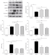
Effect of BSKS on the expression of adiponectin and its receptors as well as TLR4 and NF-κB p65 in the atherosclerotic aorta of ApoE−/− mice. The expressions of adiponectin, AdipoR1, AdipoR2, TLR4, and NF-κB p65 in the atherosclerotic aorta of ApoE−/− mice were detected by western blot. The expressions of adiponectin, AdipoR1, AdipoR2, TLR4, and NF-κB p65 were quantitatively compared between drug treatment groups and model group (n = 3). Data are shown as mean ± standard deviation and compared by one-way analysis of variance followed by Fisher's Least Significant Difference test for individual comparisons. n.s.P > 0.05; P < 0.05; ⋆⋆P < 0.01.
Figure 4
Figure 4
Effect of BSKS on the inflammatory mediators in blood of ApoE−/− mice. The serum levels of proinflammatory mediators, including IL-1β, MCP-1, and VCAM-1, and anti-inflammatory mediators, including IL-10 and adiponectin, were detected by enzyme-linked immunosorbent assay and quantitatively compared between drug treatment groups and model group (n = 10) at 6 weeks or 12 weeks. Data are shown as mean ± standard deviation and compared by one-way analysis of variance followed by Fisher's Least Significant Difference test for individual comparisons. ⋆⋆P < 0.01.

Similar articles

Cited by

References

    1. Ross R. Atherosclerosis—an inflammatory disease. The New England Journal of Medicine. 1999;340(2):115–126. doi: 10.1056/NEJM199901143400207. - DOI - PubMed
    1. Smith S. R., Lovejoy J. C., Greenway F., et al. Contributions of total body fat, abdominal subcutaneous adipose tissue compartments, and visceral adipose tissue to the metabolic complications of obesity. Metabolism - Clinical and Experimental. 2001;50(4):425–435. doi: 10.1053/meta.2001.21693. - DOI - PubMed
    1. Hansen T., Ahlström H., Söderberg S., et al. Visceral adipose tissue, adiponectin levels and insulin resistance are related to atherosclerosis as assessed by whole-body magnetic resonance angiography in an elderly population. Atherosclerosis. 2009;205(1):163–167. doi: 10.1016/j.atherosclerosis.2008.11.007. - DOI - PubMed
    1. Beltowski J. Leptin and atherosclerosis. Atherosclerosis. 2006;189(1):47–60. doi: 10.1016/j.atherosclerosis.2006.03.003. - DOI - PubMed
    1. Hopkins T. A., Ouchi N., Shibata R., Walsh K. Adiponectin actions in the cardiovascular system. Cardiovascular Research. 2007;74(1):11–18. doi: 10.1016/j.cardiores.2006.10.009. - DOI - PMC - PubMed

LinkOut - more resources