

Aptamer-conjugated nanomaterials for specific cancer cell recognition and targeted cancer therapy
- PMID: 29619132
- PMCID: PMC5880215
- DOI: 10.1038/am.2014.12
Aptamer-conjugated nanomaterials for specific cancer cell recognition and targeted cancer therapy
Abstract
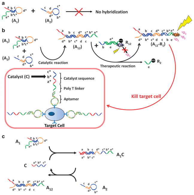
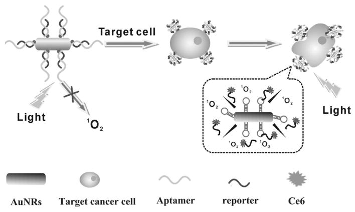
Based on their unique advantages, increasing interest has been shown in the use of aptamers as target ligands for specific cancer cell recognition and targeted cancer therapy. Recently, the development of aptamer-conjugated nanomaterials has offered new therapeutic opportunities for cancer treatment with better efficacy and lower toxicity. We highlight some of the promising classes of aptamer-conjugated nanomaterials for the specific recognition of cancer cells and targeted cancer therapy. Recent developments in the use of novel strategies that enable sensitive and selective cancer cell recognition are introduced. In addition to targeted drug delivery for chemotherapy, we also review how aptamer-conjugated nanomaterials are being incorporated into emerging technologies with significant improvement in efficiency and selectivity in cancer treatment.
Keywords: aptamer; cancer therapy; cell recognition; nanomaterials.
Conflict of interest statement
CONFLICT OF INTEREST The authors declare no conflict of interest.
Figures
Similar articles
-
Cell-specific aptamers and their conjugation with nanomaterials for targeted drug delivery.Expert Opin Drug Deliv. 2015 Mar;12(3):493-506. doi: 10.1517/17425247.2015.966681. Epub 2014 Nov 28. Expert Opin Drug Deliv. 2015. PMID: 25430795 Review.
-
Targeted cancer drug delivery with aptamer-functionalized polymeric nanoparticles.J Drug Target. 2019 Mar;27(3):292-299. doi: 10.1080/1061186X.2018.1491978. Epub 2018 Jul 26. J Drug Target. 2019. PMID: 29929413 Review.
-
Revolutionizing biomedicine: Aptamer-based nanomaterials and nanodevices for therapeutic applications.Biotechnol Rep (Amst). 2024 May 24;42:e00843. doi: 10.1016/j.btre.2024.e00843. eCollection 2024 Jun. Biotechnol Rep (Amst). 2024. PMID: 38881649 Free PMC article. Review.
-
Application of aptamer functionalized nanomaterials in targeting therapeutics of typical tumors.Front Bioeng Biotechnol. 2023 Feb 16;11:1092901. doi: 10.3389/fbioe.2023.1092901. eCollection 2023. Front Bioeng Biotechnol. 2023. PMID: 36873354 Free PMC article. Review.
-
Aptamer-Based Targeted Drug Delivery Systems: Current Potential and Challenges.Curr Med Chem. 2020;27(13):2189-2219. doi: 10.2174/0929867325666181008142831. Curr Med Chem. 2020. PMID: 30295183 Review.
Cited by
-
Multi-functional stretchable sensors based on a 3D-rGO wrinkled microarchitecture.Nanoscale Adv. 2019 Sep 27;1(11):4406-4414. doi: 10.1039/c9na00429g. eCollection 2019 Nov 5. Nanoscale Adv. 2019. PMID: 36134427 Free PMC article.
-
An enzymolysis-induced energy transfer co-assembled system for spontaneously recoverable supramolecular dynamic memory.Chem Sci. 2024 Jun 13;15(28):11084-11091. doi: 10.1039/d4sc02756f. eCollection 2024 Jul 17. Chem Sci. 2024. PMID: 39027284 Free PMC article.
-
Aptamer-Enabled Nanomaterials for Therapeutics, Drug Targeting and Imaging.Cells. 2022 Jan 4;11(1):159. doi: 10.3390/cells11010159. Cells. 2022. PMID: 35011722 Free PMC article. Review.
-
Phospholipase A2-Responsive Phosphate Micelle-Loaded UCNPs for Bioimaging of Prostate Cancer Cells.Sci Rep. 2017 Nov 22;7(1):16073. doi: 10.1038/s41598-017-16136-4. Sci Rep. 2017. PMID: 29167526 Free PMC article.
-
Harnessing molecular recognition for localized drug delivery.Adv Drug Deliv Rev. 2021 Mar;170:238-260. doi: 10.1016/j.addr.2021.01.008. Epub 2021 Jan 20. Adv Drug Deliv Rev. 2021. PMID: 33484737 Free PMC article. Review.
References
-
- Mukerjee A, Ranjan AP, Vishwanatha JK. Combinatorial nanoparticles for cancer diagnosis and therapy. Curr Med Chem. 2012;19:3714–3721. - PubMed
-
- Barbas AS, Mi J, Clary BM, White RR. Aptamer applications for targeted cancer therapy. Future Oncol. 2010;6:1117–1126. - PubMed
-
- Bamrungsap S, Zhao Z, Chen T, Wang L, Li C, Fu T, Tan W. Nanotechnology in therapeutics: a focus on nanoparticles as a drug delivery system. Nanomedicine. 2012;7:1253–1271. - PubMed
-
- Gu FX, Karnik R, Wang AZ, Alexis F, Levy-Nissenbaum E, Hong S, Langer RS, Farokhzad OC. Targeted nanoparticles for cancer therapy. Nanotoday. 2007;2:14–21.
Grants and funding
LinkOut - more resources
Full Text Sources
Other Literature Sources