. 2018 Mar 30:6:9.
doi: 10.1038/s41413-018-0012-0. eCollection 2018.

Omentin-1 prevents inflammation-induced osteoporosis by downregulating the pro-inflammatory cytokines

Affiliations

Omentin-1 prevents inflammation-induced osteoporosis by downregulating the pro-inflammatory cytokines

Shan-Shan Rao et al. Bone Res. .

Abstract

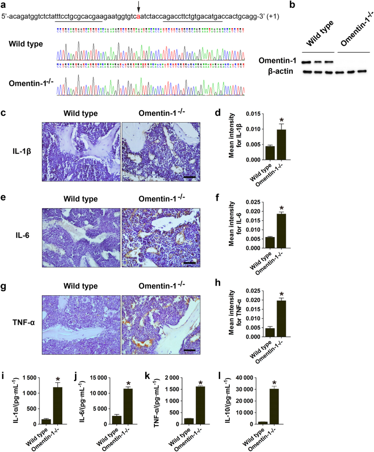
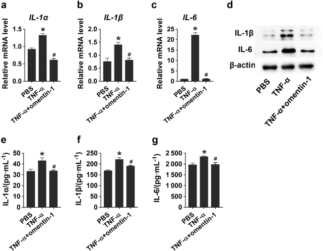
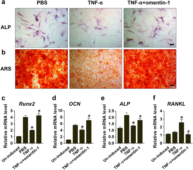
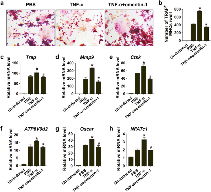
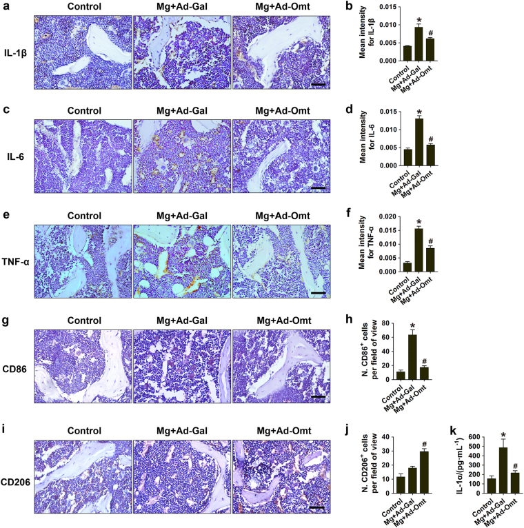
Osteoporosis is a frequent complication of chronic inflammatory diseases and increases in the pro-inflammatory cytokines make an important contribution to bone loss by promoting bone resorption and impairing bone formation. Omentin-1 is a newly identified adipocytokine that has anti-inflammatory effects, but little is known about the role of omentin-1 in inflammatory osteoporosis. Here we generated global omentin-1 knockout (omentin-1-/-) mice and demonstrated that depletion of omentin-1 induces inflammatory bone loss-like phenotypes in mice, as defined by abnormally elevated pro-inflammatory cytokines, increased osteoclast formation and bone tissue destruction, as well as impaired osteogenic activities. Using an inflammatory cell model induced by tumor necrosis factor-α (TNF-α), we determined that recombinant omentin-1 reduces the production of pro-inflammatory factors in the TNF-α-activated macrophages, and suppresses their anti-osteoblastic and pro-osteoclastic abilities. In the magnesium silicate-induced inflammatory osteoporosis mouse model, the systemic administration of adenoviral-delivered omentin-1 significantly protects from osteoporotic bone loss and inflammation. Our study suggests that omentin-1 can be used as a promising therapeutic agent for the prevention or treatment of inflammatory bone diseases by downregulating the pro-inflammatory cytokines.


Conflict of interest statement

The authors declare that they have no conflict of interest.

Figures

Fig. 1
Fig. 1
Omentin-1 depletion induces inflammatory responses in mice. a, b One base (a) was inserted into the sequence of omentin-1 gene on exon 4 in one strand, resulting in the early termination of omentin-1 protein translation. Genomic DNA from tail tips of omentin-1−/− mice and their wild-type littermates was genotyped by DNA sequencing (a). The depletion of omentin-1 in omentin-1−/− mice was verified by western blot analysis of omentin-1 protein in the small intestines from omentin-1−/− mice and their wild-type littermates. n = 3 per group (b). ch Representative images of immunohistochemical staining for IL-1β (n = 6 or 9 per group), IL-6 (n = 4 or 6 per group) and TNF-α (n = 6 or 9 per group) in femora of omentin-1−/− and wild-type mice. The graphs show mean intensity for the positively stained areas. Scale bar: 50 μm. il The serum concentrations of pro-inflammatory cytokines IL-1α, IL-6, and TNF-α (ik) and the anti-inflammatory factor IL-10 (l) in omentin-1−/− and wild-type mice, as determined by ELISA. n = 6 or 9 per group. Data are shown as the mean ± s.d. *P < 0.05 compared with the control (wild-type) mice.
Fig. 2
Fig. 2
Omentin-1 depletion leads to abnormal bone loss in mice. a Representative microcomputed tomography (µCT) images of femora in omentin-1−/− and wild-type mice. Scale bar: 1 mm. bg Quantitative analyses of the trabecular bone fraction (Tb.BV/TV), trabecular thickness (Tb.Th), trabecular number (Tb.N), trabecular separation (Tb.Sp), periosteal perimeter (Ps.Pm) and cortical bone thickness (Ct.Th) of femora. n = 6 per group. h Representative images of immunohistochemical staining for OCN. Scale bar: 50 μm. i Quantitative analysis of the mean intensity for the positively stained areas in h. n = 5 or 6 per group. j ELISA of the serum concentration of OCN. n = 5 or 7 per group. k TRAP staining of femora in omentin-1−/− and wild-type mice. Scale bar: 50 μm. l Quantitative analysis of the number of TRAP+ cells in k. n = 5 or 6 per group. BS bone surface, OCs osteoclasts. m ELISA of the serum concentration of CTX-I. n = 10 per group. Data are shown as the mean ± s.d. *P < 0.05 compared with the wild-type mice.
Fig. 3
Fig. 3
Omentin-1 impairs TNF-α-induced inflammation in macrophages. ac RAW264.7 macrophages were treated with 10 ng·mL-1 TNF-α or an equal volume diluents (PBS) for 30 min. The culture medium was replaced to remove residual TNF-α and the cells were then incubated in fresh medium supplemented with 300 μg·mL-1 omentin-1 or PBS for 24 h. The mRNA levels of IL-1α, IL-1β, and IL-6 in RAW264.7 macrophages receiving different treatments were tested by qRT-PCR analysis. n = 3 per group. d Detection of protein levels of IL-1β and IL-6 in macrophages receiving different treatments. eg The concentrations of IL-1α, IL-1β, and IL-6 in conditioned media (CM) derived from macrophages receiving different treatments. n = 3 per group. All the results are representative of three independent experiments. Data are shown as the mean ± s.d. *P < 0.05 compared with the PBS group, #P < 0.05 compared with the TNF-α group.
Fig. 4
Fig. 4
Omentin-1 inhibits the anti-osteoblastic effects of TNF-α-activated macrophages. a, b MSCs were cultured in osteogenic medium supplemented with the CM from macrophages treated with PBS (30 min + 24 h), TNF-α (10 ng·mL-1; 30 min), or TNF-α (10 ng·mL-1; 30 min) + omentin-1 (300 μg·mL-1; 24 h). Alkaline phosphatase (ALP) (a) and alizarin red S (ARS) staining (b) were performed after 3 and 14 days of induction, respectively. n = 3 per group. Scale bar: 100 μm. cf The expression levels of Runx2, OCN, ALP, and RANKL were detected by qRT-PCR analyses on day 7. n = 3 per group. All the results are representative of three independent experiments. Data are shown as the mean ± s.d. *P < 0.05 compared with the PBS group (cells treated with CM from inactivated macrophages), #P < 0.05 compared with the TNF-α group (cells treated with CM from TNF-α-activated macrophages).
Fig. 5
Fig. 5
Omentin-1 downregulates the pro-osteoclastic abilities of TNF-α-activated macrophages. a TRAP staining of RAW264.7 cells cultured in RANKL-containing medium supplemented with CM from macrophages treated with PBS (30 min + 24 h), TNF-α (10 ng·mL-1; 30 min), or TNF-α (10 ng·mL-1; 30 min) + omentin-1 (300 μg·mL-1; 24 h). Scale bar: 50 μm. b Quantitative analysis of the number of TRAP+ cells in a. n = 3 per group. ch The expression levels of osteoclastogenesis-related genes including Trap, Mmp9, Ctsk, ATP6V0d2, Oscar, and NFATc1 were assessed by qRT-PCR analyses. n = 3 per group. All the results are representative of three independent experiments. Data are shown as the mean ± s.d. *P < 0.05 compared with the PBS group (cells treated with CM from inactivated macrophages), #P < 0.05 compared with the TNF-α group (cells treated with CM from TNF-α-activated macrophages).
Fig. 6
Fig. 6
Omentin-1 attenuates inflammatory responses induced by magnesium silicate. ac Immunohistochemical staining of IL-1β, IL-6, and TNF-α in bone marrow and on bone surface in mice receiving different treatments. Scale bar: 50 μm. df Quantitative analyses of the mean intensity of the positively stained areas in ac. n = 5–7 per group. g Immunohistochemical staining for CD86 (a marker for pro-inflammatory M1 macrophages). Scale bar: 50 μm. h Quantitative analysis of the numbers of CD86+ cells in g. n = 3 per group. i Immunohistochemical staining for CD206 (a marker for anti-inflammatory M2 macrophages). Scale bar: 50 μm. j Quantitative analysis of the numbers of CD206+ cells in i. n = 3 per group. k The serum concentration of IL-1α in different treatment groups, as analyzed by ELISA. n = 5–9 per group. Data are shown as the mean ± s.d. *P < 0.05 compared with the control group, #P < 0.05 compared with the magnesium silicate + Ad-Gal (Mg + Ad-Gal) group.
Fig. 7
Fig. 7
Omentin-1 prevents magnesium silicate-induced inflammatory bone loss. a Representative µCT images of femora in mice receiving different treatments. Scale bar: 1 mm. bg Quantitative analyses of Tb.BV/TV, Tb.Th, Tb.N, Tb.Sp, Ps.Pm, and Ct.Th of femora in a. n = 6–8 per group. h Representative images of immunohistochemical staining for OCN. Scale bar: 50 μm. i Quantitative analysis of the mean intensity of OCN positively stained areas in h. n = 5–6 per group. j ELISA of the serum concentration of OCN. n = 6–8 per group. k TRAP staining of femora in mice receiving different treatments. Scale bar: 50 μm. l Quantitative analysis of the number of TRAP+ cells in k. n = 6–7 per group. m ELISA of the serum concentration of CTX-I. n = 6–7 per group. The data are shown as the mean ± s.d. *P < 0.05 compared with the control group, #P < 0.05 compared with the Mg + Ad-Gal group.

References

    1. Amarasekara DS, Yu J, Rho J. Bone loss triggered by the cytokine network in inflammatory autoimmune diseases. J. Immunol. Res. 2015;2015:832127. doi: 10.1155/2015/832127. - DOI - PMC - PubMed
    1. Lv L, et al. Lysine-specific demethylase 1 inhibitor rescues the osteogenic ability of mesenchymal stem cells under osteoporotic conditions by modulating h3k4 methylation. Bone Res. 2016;4:16037. doi: 10.1038/boneres.2016.37. - DOI - PMC - PubMed
    1. Duan X, et al. Deficiency of atp6v1h causes bone loss by inhibiting bone resorption and bone formation through the tgf-beta1 pathway. Theranostics. 2016;6:2183–2195. doi: 10.7150/thno.17140. - DOI - PMC - PubMed
    1. Baum R, Gravallese EM. Impact of inflammation on the osteoblast in rheumatic diseases. Curr. Osteoporos. Rep. 2014;12:9–16. doi: 10.1007/s11914-013-0183-y. - DOI - PMC - PubMed
    1. Pan HY, Guo L, Li Q. Changes of serum omentin-1 levels in normal subjects and in patients with impaired glucose regulation and with newly diagnosed and untreated type 2 diabetes. Diabetes Res. Clin. Pract. 2010;88:29–33. doi: 10.1016/j.diabres.2010.01.013. - DOI - PubMed