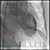
Takotsubo cardiomyopathy with Basal Hypertrophy and outflow obstruction in a patient with bowel ischemia
- PMID: 29619340
- PMCID: PMC5869801
- DOI: 10.4103/IJCIIS.IJCIIS_47_17
Takotsubo cardiomyopathy with Basal Hypertrophy and outflow obstruction in a patient with bowel ischemia
Abstract
Basal septal hypertrophy is a rare and unique anatomical finding associated with hypertrophic cardiomyopathy (HCM). It is also described as a sigmoid hypertrophy and is linked with aging and chronic hypertension. Takotsubo cardiomyopathy is a transient cardiomyopathy that occurs during periods of high physical or emotional stress. Its occurrence with HCM is relatively common; however, this presentation occurs more often with the classic asymmetrical septal hypertrophy or the apical variant. This case demonstrates its coexistence with isolated sigmoid hypertrophy in an elderly, hypertensive female with severe ischemic bowel disease.
Keywords: Basal hypertrophy; bowel ischemia; takotsubo.
Conflict of interest statement
There are no conflicts of interest.
Figures


Similar articles
-
An unusual case of severe left ventricle outflow tract obstruction due to a coexistence of Takotsubo cardiomyopathy with septal hypertrophic cardiomyopathy.Monaldi Arch Chest Dis. 2021 Dec 30;92(3). doi: 10.4081/monaldi.2021.2106. Monaldi Arch Chest Dis. 2021. PMID: 35658329
-
Syncope in a hypertrophic heart at a wedding party: can happiness break a thick heart? Takotsubo cardiomyopathy complicated with left ventricular outflow tract obstruction in a hypertrophic heart.Oxf Med Case Reports. 2020 Jun 25;2020(6):omaa036. doi: 10.1093/omcr/omaa036. eCollection 2020 Jun. Oxf Med Case Reports. 2020. PMID: 32626581 Free PMC article.
-
Surgical pathology of subaortic septal myectomy: histology skips over clinical diagnosis.Cardiovasc Pathol. 2018 Mar-Apr;33:32-38. doi: 10.1016/j.carpath.2017.12.002. Epub 2018 Jan 3. Cardiovasc Pathol. 2018. PMID: 29414430
-
Surgical treatment of hypertrophic cardiomyopathy.Expert Rev Cardiovasc Ther. 2013 May;11(5):617-27. doi: 10.1586/erc.13.46. Expert Rev Cardiovasc Ther. 2013. PMID: 23621143 Review.
-
Hypertrophic cardiomyopathy.Cardiol Clin. 1988 May;6(2):233-88. Cardiol Clin. 1988. PMID: 3066484 Review.
References
-
- Armstrong WF, Ryan T, Feigenbaum H. Feigenbaum's Echocardiography. 7th ed. Philadelphia: Wolters Kluwer Health/Lippincott Williams & Wilkins; 2010.
-
- Turer AT, Samad Z, Valente AM, Parker MA, Hayes B, Kim RJ, et al. Anatomic and clinical correlates of septal morphology in hypertrophic cardiomyopathy. Eur J Echocardiogr. 2011;12:131–9. - PubMed
-
- Aboulhosn J, Child JS. Left ventricular outflow obstruction: Subaortic stenosis, bicuspid aortic valve, supravalvar aortic stenosis, and coarctation of the aorta. Circulation. 2006;114:2412–22. - PubMed
Publication types
LinkOut - more resources
Full Text Sources
Other Literature Sources

