

The Use of Ultrasound Imaging in the External Beam Radiotherapy Workflow of Prostate Cancer Patients
- PMID: 29619375
- PMCID: PMC5829356
- DOI: 10.1155/2018/7569590
The Use of Ultrasound Imaging in the External Beam Radiotherapy Workflow of Prostate Cancer Patients
Abstract
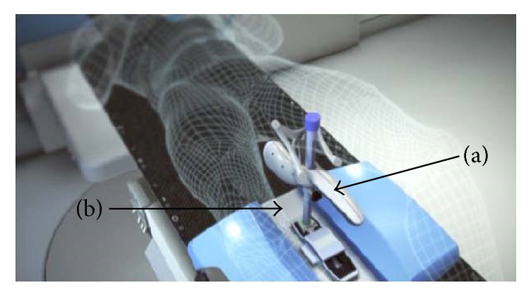
External beam radiotherapy (EBRT) is one of the curative treatment options for prostate cancer patients. The aim of this treatment option is to irradiate tumor tissue, while sparing normal tissue as much as possible. Frequent imaging during the course of the treatment (image guided radiotherapy) allows for determination of the location and shape of the prostate (target) and of the organs at risk. This information is used to increase accuracy in radiation dose delivery resulting in better tumor control and lower toxicity. Ultrasound imaging is harmless for the patient, it is cost-effective, and it allows for real-time volumetric organ tracking. For these reasons, it is an ideal technique for image guidance during EBRT workflows. Review papers have been published in which the use of ultrasound imaging in EBRT workflows for different cancer sites (prostate, breast, etc.) was extensively covered. This new review paper aims at providing the readers with an update on the current status for prostate cancer ultrasound guided EBRT treatments.
Figures
References
-
- Fitzmaurice C., Allen C., Barber R. M., et al. Global, regional, and national cancer incidence, mortality, years of life lost, years lived with disability, and disability-adjusted life-years for 32 cancer groups, 1990 to 2015: a systematic analysis for the global burden of disease study. JAMA Oncology. 2017;3(4):524–548. - PMC - PubMed
-
- Zelefsky M. J., Kollmeier M., Cox B., et al. Improved clinical outcomes with high-dose image guided radiotherapy compared with non-IGRT for the treatment of clinically localized prostate cancer. International Journal of Radiation Oncology • Biology • Physics. 2012;84(1):125–129. doi: 10.1016/j.ijrobp.2011.11.047. - DOI - PubMed
-
- Sveistrup J., af Rosenschöld P. M., Deasy J. O., et al. Improvement in toxicity in high risk prostate cancer patients treated with image-guided intensity-modulated radiotherapy compared to 3D conformal radiotherapy without daily image guidance. Journal of Radiation Oncology. 2014;9(1, article 44) doi: 10.1186/1748-717x-9-44. - DOI - PMC - PubMed
Publication types
MeSH terms
LinkOut - more resources
Full Text Sources
Other Literature Sources
Medical
