Review
. 2018 Apr 5:4:18016.
doi: 10.1038/nrdp.2018.16.

Haemochromatosis

Affiliations
Review

Haemochromatosis

Pierre Brissot et al. Nat Rev Dis Primers. .

Abstract

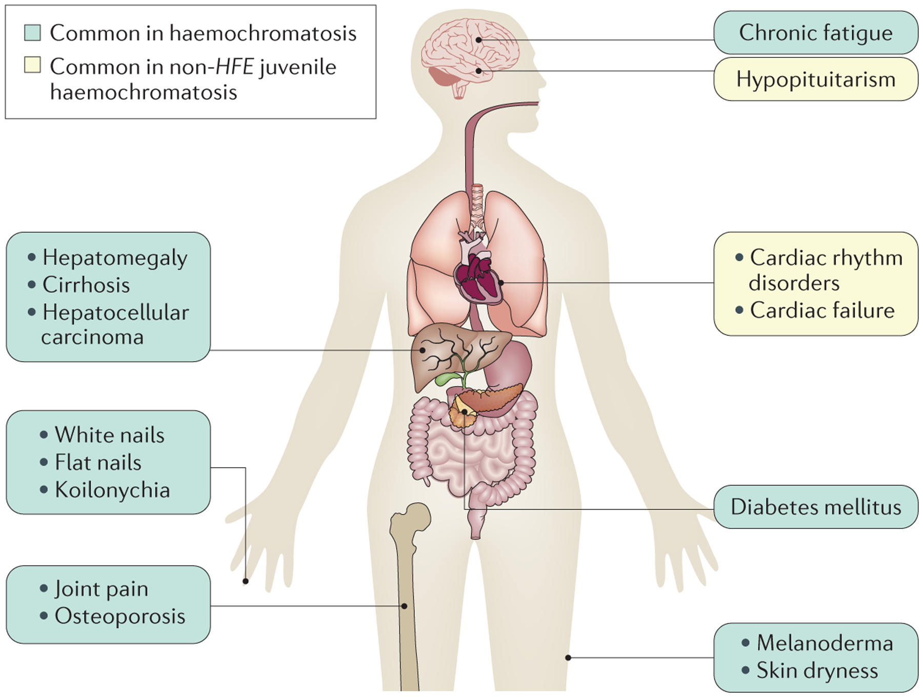
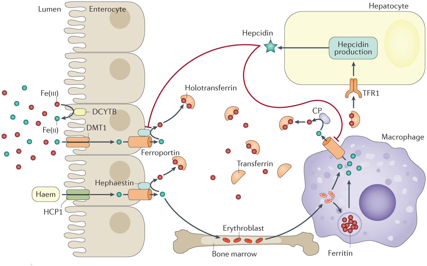
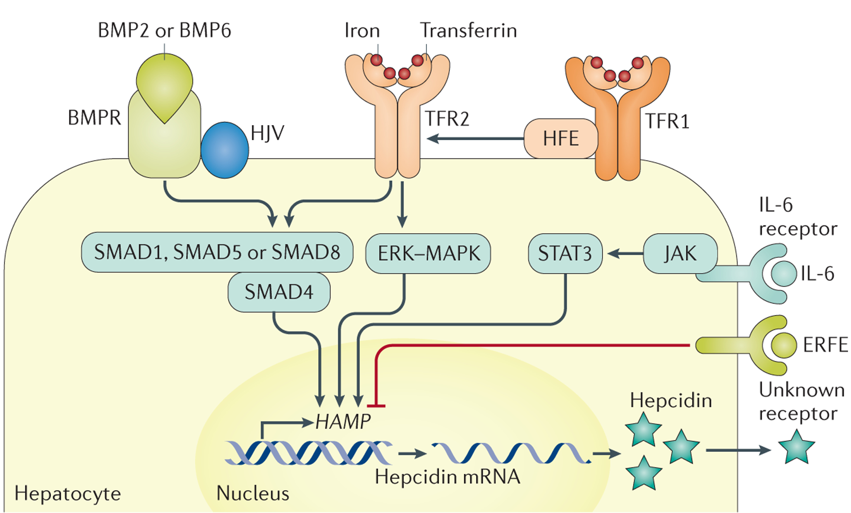
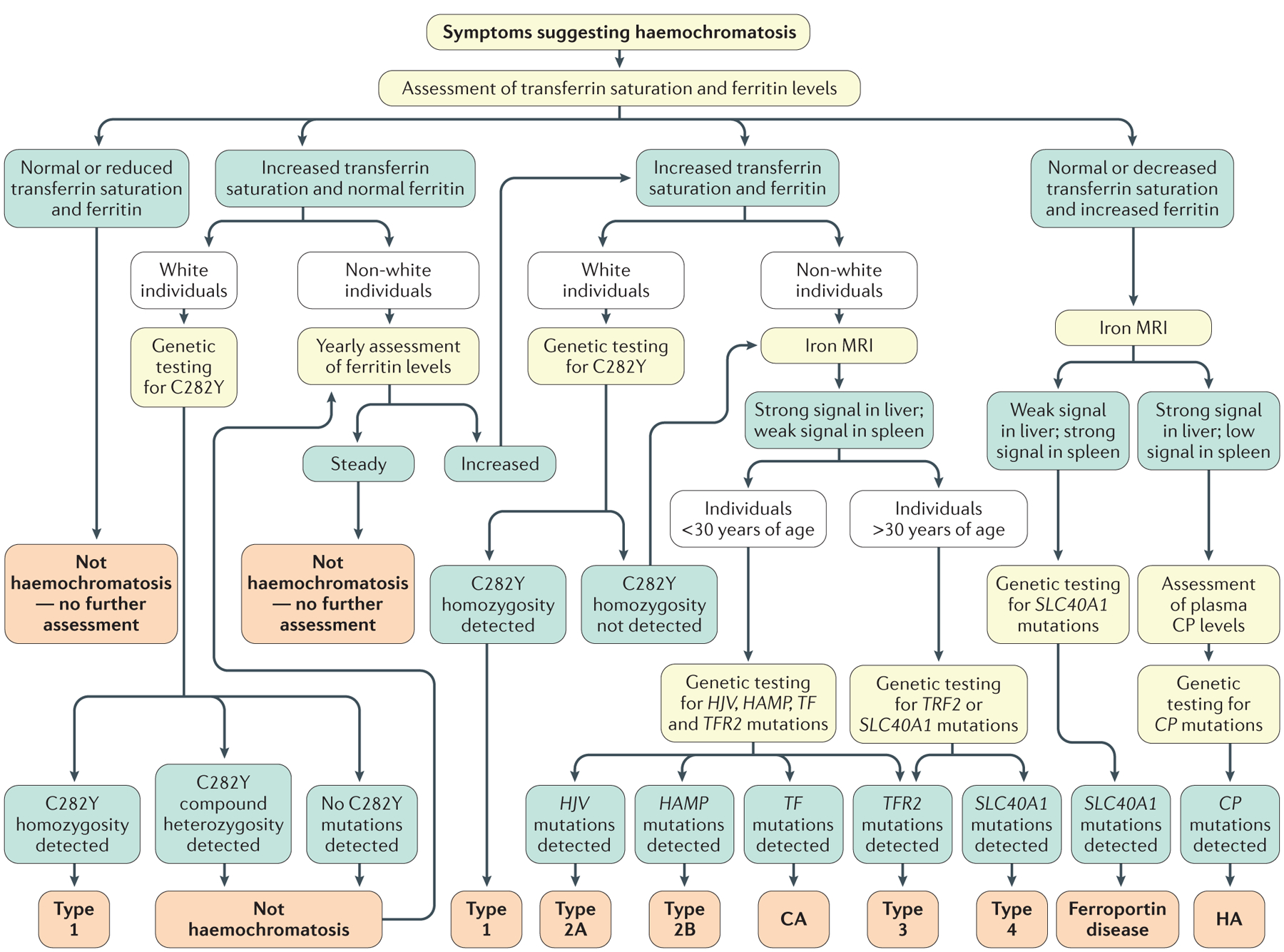
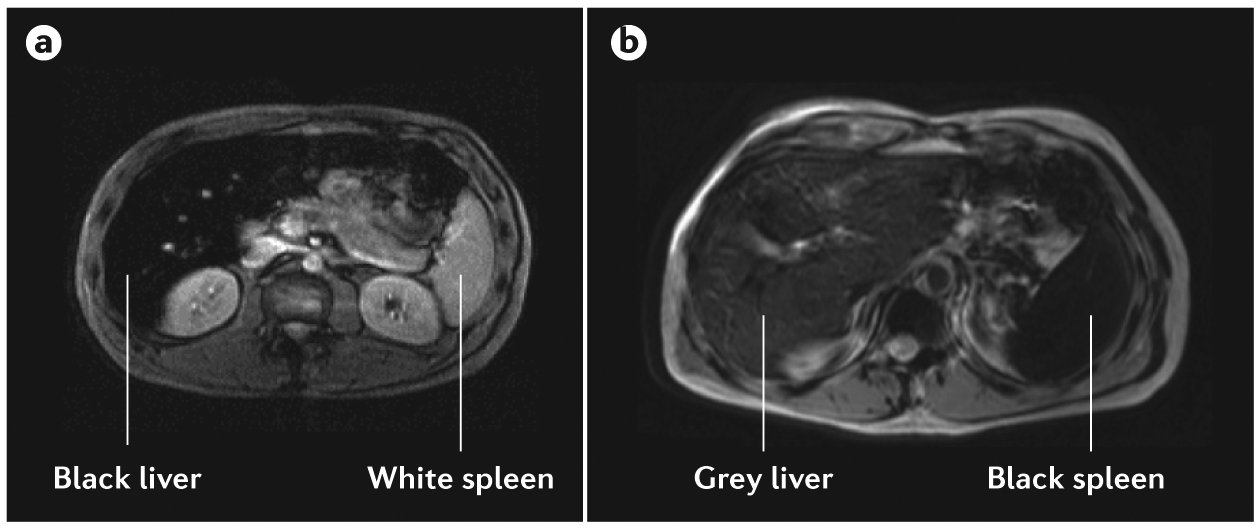
Haemochromatosis is defined as systemic iron overload of genetic origin, caused by a reduction in the concentration of the iron regulatory hormone hepcidin, or a reduction in hepcidin-ferroportin binding. Hepcidin regulates the activity of ferroportin, which is the only identified cellular iron exporter. The most common form of haemochromatosis is due to homozygous mutations (specifically, the C282Y mutation) in HFE, which encodes hereditary haemochromatosis protein. Non-HFE forms of haemochromatosis due to mutations in HAMP, HJV or TFR2 are much rarer. Mutations in SLC40A1 (also known as FPN1; encoding ferroportin) that prevent hepcidin-ferroportin binding also cause haemochromatosis. Cellular iron excess in HFE and non-HFE forms of haemochromatosis is caused by increased concentrations of plasma iron, which can lead to the accumulation of iron in parenchymal cells, particularly hepatocytes, pancreatic cells and cardiomyocytes. Diagnosis is noninvasive and includes clinical examination, assessment of plasma iron parameters, imaging and genetic testing. The mainstay therapy is phlebotomy, although iron chelation can be used in some patients. Hepcidin supplementation might be an innovative future approach.


Conflict of interest statement

Competing interests

P.B. has received lecture fees from Novartis and consulting fees from Novartis and La Jolla Pharmaceutical Company. A.P. has received lecture fees from Novartis, and consulting fees from Novartis, La Jolla Pharmaceutical Company and Mitsubishi Tanabe Pharma Corporation. O.L. has received a research grant from Novartis. All other authors declare no competing interests.

Figures

Figure 1 |
Figure 1 |. Symptoms of haemochromatosis.
When symptoms develop, chronic fatigue is prominent. In addition, joint pain is frequent and is caused by acute or chronic monoarthritis, oligoarthritis or polyarthritis; arthritis in the second and third metacarpophalangeal joints and the ankles is particularly suggestive of haemochromatosis. Spontaneous fractures (particularly of the vertebrae) can occur owing to early-onset osteoporosis. Dermatological signs are primarily melanoderma (darkening of the skin) but can also include skin dryness and nail changes, such as white nails, flat nails and koilonychia (that is, abnormally thin nails that curve inwards, also called ‘spoon’ nails). The main hepatic symptom is hepatomegaly. In contrast to most other liver diseases (notably due to alcohol use, viral infection or nonalcoholic fatty liver), cirrhotic livers in patients with haemochromatosis are well functioning, such that neither hepatocellular insufficiency or portal hypertension are usually observed in the absence of hepatotoxic cofactors. This finding explains why patients with haemochromatosis rarely develop complications of liver failure but develop hepatocellular carcinoma. In addition, diabetes mellitus and, more rarely, adrenal insufficiency or hypopituitarism can occur. Cardiac symptoms consist of cardiac rhythm disorders and cardiac failure. Anaemic syndrome is not observed in haemochromatosis; the presence of this symptom together with signs of iron excess suggests congenital atransferrinaemia, hereditary aceruloplasminaemia and divalent cation transporter 1 (DMT1)-related iron overload. Arthropathies are a frequent complication of haemochromatosis that frequently persists in patients despite otherwise successful iron depletive treatments, suggesting that this manifestation is not directly related to iron excess. Whether persistent high plasma transferrin saturation levels with the presence of labile plasma iron are involved in arthropathies needs further investigation.
Figure 2 |
Figure 2 |. Iron uptake, cycling and distribution within the body.
The main sources of plasma iron are enterocytes and macrophages. Non-haem iron is absorbed through divalent cation transporter 1 (DMT1), which is found on the apical surface of enterocytes after conversion of iron from its ferric (Fe(iii)) to its ferrous (Fe(ii)) form by a reduction process that can involve the duodenal cytochrome b reductase 1 (CYBDR1; also known as DCYTB) or other enzymes. The mechanism of haem iron uptake by enterocytes is not fully elucidated, although it likely involves proton-coupled folate transporter (SLC46A1; also known as HCP1). By contrast, macrophages acquire iron from erythrophagocytosis (whereby erythrocytes are degraded and the contained iron is recycled). Iron is released from enterocytes and macrophages into plasma through ferroportin. Iron is then oxidized by ceruloplasmin (CP, which circulates in plasma) and hephaestin (which is anchored to enterocytes) and binds to transferrin (to form holotransferrin),. CP has a role in the control of iron export from reticuloendothelial cells, and both hephaestin and CP might have a role in cell iron metabolism in other tissues,. Holotransferrin delivers iron to every cell type, although erythroblasts (immature erythrocytes) are the main consumers. Holotransferrin levels are also sensed by hepatocytes that take up iron through transferrin receptor 1 (TFR1). In response to high levels of holotransferrin, hepcidin — which induces the degradation of ferroportin — is secreted into plasma to control the iron export. Figure adapted from REF. , Macmillan Publishers Limited.
Figure 3 |
Figure 3 |. Hepcidin regulation.
Increased cell iron stores lead to increased bone morphogenetic protein 6 (BMP6) expression in liver cells (sinusoidal cells are probably the major producers,, but hepatocytes and stellate cells might also be involved). BMP6 binds to the heterodimeric BMP receptor (BMPR) type 1 and type 2, which are bound to haemojuvelin (HJV),. BMP-BMPR binding leads to BMPR phosphorylation, which leads to the phosphorylation of mothers against decapentaplegic homologue 1 (SMAD1), SMAD5 or SMAD8 (REF. 221). These proteins form a complex with SMAD4, which is transported into the nucleus and interacts with BMP-responsive elements on the HAMP promoter, leading to HAMP transcription and hepcidin expression,–. Transferrin saturation is also involved in hepcidin regulation. This process could involve a shift of the interactions between hereditary haemochromatosis protein (HFE), transferrin receptor 2 (TFR2) and transferrin receptor 1 (TFR1) with increased transferrin saturation, leading to signalling — potentially through mitogen-activated protein kinase (MAPK) and extracellular-signal-regulated kinase (ERK) — to increase HAMP transcription, although this process is not fully understood. The HFE-TFR1-TFR2 complex could also interact with the BMPR-HJV complex. Other regulators of hepcidin expression include chronic inflammation, which is mediated by IL-6 produced by inflammatory cells and induces Janus kinase (JAK) and signal transducers and activators of transcription 3 (STAT3) activation, in addition to erythroferrone (ERFE), which is produced by erythroblasts and interacts with unknown partners to decrease HAMP transcription. Figure adapted from REF. , Macmillan Publishers Limited.
Figure 4 |
Figure 4 |. Diagnostic chart for systemic iron overload of genetic origin.
This algorithm excludes the diagnosis of acquired iron overload owing to blood transfusions, dyserythropoiesis or parenteral iron supplementation. Several factors should be considered as part of the initial diagnostic workup for haemochromatosis, such as patient ethnicity (as HFE-associated haemochromatosis is observed almost exclusively in white individuals) and sex (as the phenotypic expression of haemochromatosis is usually less pronounced in women). In addition, patient age should be taken into consideration, as HFE-associated (type 1) or TFR2-associated (type 3) haemochromatosis is generally observed in individuals >30 years of age, whereas clinical expression in younger individuals is typical of HJV-related (type 2A) or HAMP-related (type 2B) haemochromatosis. The diagnostic approach should also consider that non-HFE haemochromatosis diseases are rare, in contrast to HFE-associated haemochromatosis. Type 4 refers to haemochromatosis caused by gain-of-function mutations in SLC40A1, previously named haemochromatosis type 4B. CA, congenital atransferrinaemia; CP, ceruloplasmin; HA, hereditary aceruloplasminaemia.
Figure 5 |
Figure 5 |. MRI findings in haemochromatosis owing to hepcidin deficiency and ferroportin disease.
The signal intensity ratio technique with T2-weighted MRI can be used to differentiate patients with haemochromatosis and those with ferroportin disease. a | In patients with hepcidin-deficient haemochromatosis, ‘black’ liver corresponds to a highly iron-overloaded liver, and ‘white’ spleen corresponds to the absence of iron overload. The appearance of the liver and spleen is similar in patients with types 1, 2A, 2B, 3 and 4 haemochromatosis. b | In ferroportin disease, the spleen appears black (highly iron-overloaded) and the liver appears grey (moderately iron-overloaded) or black (highly iron-overloaded) on T2-weighted MRI.
Figure 6 |
Figure 6 |. Proposed phenotypic classification of haemochromatosis related to hepcidin deficiency.
Increased ferritin: ≥300 μg per litre for men and ≥200 μg per litre for women; increased transferrin saturation (TS): >45% (often 60–100%). QOL, quality of life. Adapted with permission from REF. , Elsevier.

References

    1. Merryweather-Clarke AT, Pointon JJ, Jouanolle AM, Rochette J & Robson KJ Geography of HFE C282Y and H63D mutations. Genet. Test 4, 183–198 (2000). - PubMed
    1. McLaren CE et al. Hemochromatosis and Iron Overload Screening (HEIRS) study design for an evaluation of 100,000 primary care-based adults. Am. J. Med. Sci 325, 53–62 (2003). - PubMed
    1. Adams PC et al. Hemochromatosis and iron-overload screening in a racially diverse population. N. Engl. J. Med 352, 1769–1778 (2005). - PubMed
    1. Kirk L et al. Haemochromatosis gene frequency in a control and diabetic Irish population. Ir. J. Med. Sci 178, 39–42 (2009). - PubMed
    1. Hanson EH, Imperatore G & Burke W HFE gene and hereditary hemochromatosis: a HuGE review. Hum. Genome Epidemiol. Am. J. Epidemiol 154, 193–206 (2001). - PubMed

Publication types