

Diagnostic and prognostic values of contrast‑enhanced ultrasound combined with diffusion‑weighted magnetic resonance imaging in different subtypes of breast cancer
- PMID: 29620140
- PMCID: PMC5979941
- DOI: 10.3892/ijmm.2018.3591
Diagnostic and prognostic values of contrast‑enhanced ultrasound combined with diffusion‑weighted magnetic resonance imaging in different subtypes of breast cancer
Retraction in
-
[Retracted] Diagnostic and prognostic value of contrast‑enhanced ultrasound combined with diffusion‑weighted magnetic resonance imaging in different subtypes of breast cancer.Int J Mol Med. 2020 Apr;45(4):1270. doi: 10.3892/ijmm.2020.4484. Epub 2020 Feb 4. Int J Mol Med. 2020. PMID: 32124936 Free PMC article.
Abstract
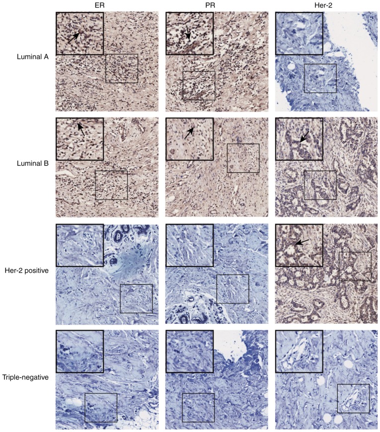
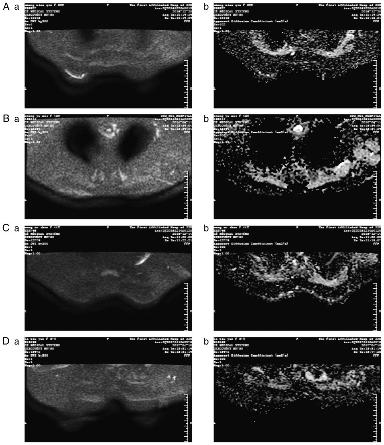
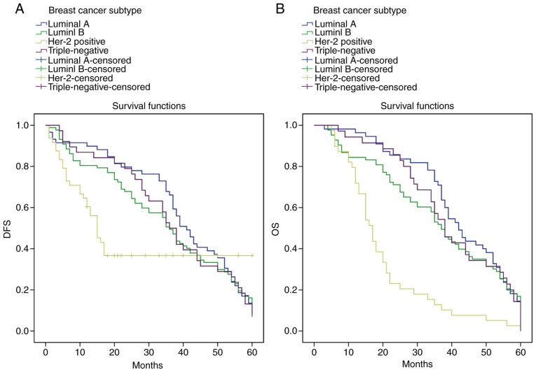
The present study aimed to investigate the diagnostic and prognostic values of contrast‑enhanced ultrasound (CEUS) combined with diffusion‑weighted magnetic resonance imaging (DW‑MRI) in different subtypes of breast cancer (BC). CEUS and DW‑MRI were conducted in 232 patients with BC prior to surgical treatment. Patients were categorized as having the luminal A subtype, the luminal B subtype, triple‑negative subtype or the human epidermal growth factor receptor 2 (Her‑2)‑positive subtype according to their expression of the estrogen receptor (ER), progesterone receptor (PR) and Her‑2, as detected by immunohistochemistry. The CEUS and DW‑MRI parameters of patients with different subtypes of BC were obtained and analyzed. The risk factors for the prognosis of patients with different subtypes of BC were analyzed using Kaplan‑Meier and COX regression analyses. The diagnostic accuracy rate of CEUS combined with DW‑MRI (93.10%) was higher than that of CEUS (88.79%) or DW‑MRI (82.33%) alone. The local recurrence rate and distant metastasis rate of the Her‑2‑positive subtype were the highest among all the subtypes. Furthermore, patients with Her‑2‑positive BC exhibited a higher proportion of lesions with indistinct margins and histological grade III. Lymph node metastasis and BC subtype were independent risk factors for the prognosis of BC. The overall survival and disease‑free survival of patients with the luminal A subtype were higher than those of patients with the Her‑2‑positive subtype. The results of the current study therefore indicate that CEUS combined with DW‑MRI is more effective at diagnosing the different subtypes of BC than either CEUS or DW‑MRI alone.
Figures
References
Publication types
MeSH terms
Substances
LinkOut - more resources
Full Text Sources
Other Literature Sources
Medical
Research Materials
Miscellaneous
