. 2018 Jul;42(1):279-289.
doi: 10.3892/ijmm.2018.3600. Epub 2018 Mar 29.

Adenovirus‑mediated knockdown of activin A receptor type 2A attenuates immune‑induced hepatic fibrosis in mice and inhibits interleukin‑17‑induced activation of primary hepatic stellate cells

Affiliations

Adenovirus‑mediated knockdown of activin A receptor type 2A attenuates immune‑induced hepatic fibrosis in mice and inhibits interleukin‑17‑induced activation of primary hepatic stellate cells

Hongjun Zhang et al. Int J Mol Med. 2018 Jul.

Abstract

Fibrosis induces a progressive loss of liver function, thus leading to organ failure. Activins are secreted proteins that belong to the transforming growth factor (TGF)‑β superfamily, which initiate signaling by binding to their two type II receptors: Activin A receptor type 2A (ACVR2A) and activin A receptor type 2B. Previous studies that have explored the mechanisms underlying immune‑induced hepatic fibrosis have mainly focused on TGF‑β signaling, not activin signaling. To investigate the role of the activin pathway in this disease, adenovirus particles containing short hairpin (sh)RNA targeting ACVR2A mRNA (Ad‑ACVR2A shRNA) were administered to mice, which were chronically treated with concanavalin A (Con A). The pathological changes in the liver were evaluated with hematoxylin/eosin staining, Masson trichrome staining and immunohistochemical assay. The results detected an increase in serum activin A and liver ACVR2A in Con A‑treated animals. Conversely, liver function was partially restored and fibrotic injury was attenuated when activin signaling was blocked. In addition, the activation of hepatic stellate cells (HSCs) in response to Con A was suppressed by Ad‑ACVR2A shRNA, as evidenced by decreased α‑smooth muscle actin, and type I and IV collagen expression. Furthermore, primary mouse HSCs (mHSCs) were activated when exposed to interleukin (IL)‑17A or IL‑17F, which are two major cytokines produced by cluster of differentiation 4+ T helper 17 cells. The levels of activin A, type I and IV collagen were determined with ELISA kits and the expression of fibrotic molecules was determined with western blot analysis. Conversely, blocking activin/ACVR2A impaired the potency of HSCs to produce collagens in response to IL‑17s. In addition, C terminus phosphorylation of Smad2 on Ser465 and Ser467, induced by either Con A in the liver or by IL‑17s in mHSCs, was partly inhibited when activin A/ACVR2A signaling was suppressed. Collectively, the present study demonstrated an involvement of activated activin A/ACVR2A/Smad2 signaling in immune‑induced hepatic fibrosis.


Figures

Figure 1
Figure 1
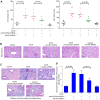
Chronic treatment with Con A induces activation of activin and IL-17 signaling in mouse liver. (A) Experiment part 1: Mice were administered Con A (8 mg/kg/week) for up to 6 weeks, in order to generate a model of immune-associated liver fibrosis. (B–E) Serum and liver levels of activin A, IL-17A and IL-17F were detected in mice in the control and Con A groups using specific ELISA kits. (F) mRNA and (G) protein expression levels of ACVR2A were determined using reverse transcription-quantitative polymerase chain reaction and western blot analysis, respectively. Data are expressed as the means ± standard deviation. (H) Experiment part 2: Mice in the Con A groups were administered two injections of Ad-ACVR2A shRNA or Ad-NC shRNA at the indicated timepoints. (I–K) Protein expression levels of ACVR2A were analyzed in mouse liver tissues collected from the various groups. Data are expressed as the means ± standard error. ACVR2A, activin A receptor type 2A; Con A, concanavalin A; IL-17, interleukin-17; NC, negative control; shRNA, short hairpin RNA.
Figure 2
Figure 2
Knockdown of ACVR2A improves liver function and attenuates fibrosis in Con A-treated mice. (A) Activity of liver function indices ALT and AST in serum samples were determined using commercial detection kits (n=7-8/group). (B) H&E staining was applied to detect the morphological structure of the liver. (C) Hepatic fibrosis was determined using the Masson trichrome assay (fibrotic area, blue). (D) Hydroxyproline content was assessed using a commercial kit. Data are expressed as the means ± standard deviation (n=8/group). Scale bars, 100 μm. ACVR2A, activin A receptor type 2A; ALT, alanine aminotransferase; AST, aspartate aminotransferase; Con A, concanavalin A; H&E, hematoxylin and eosin; NC, negative control; shRNA, short hairpin RNA.
Figure 3
Figure 3
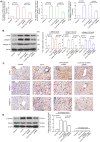
Knockdown of ACVR2A suppresses Con A-induced activation of hepatic stellate cells in vivo. (A) Relative mRNA and (B) protein expression levels of ACTA2, COL1A1 and COL4A1 were determined using reverse transcription-quantitative polymerase chain reaction and western blot analysis, respectively. (C) Immunohistochemistry was performed to detect the protein expression levels of α-SMA, collagen I and IV. Orange arrowheads, representative α-SMA-positive cells; pink arrowheads, representative collagen I-positive cells; green arrowheads, representative collagen IV-positive cells. Scale bars, 50 μm. (D) Protein expression levels of p-Smad2 and total Smad2 in liver tissues were detected using western blot analysis. Data are expressed as the means ± standard error (n=5/group). α-SMA, α-smooth muscle actin; ACVR2A, activin A receptor type 2A; Con A, concanavalin A; ; NC, negative control; p-, phosphorylated; shRNA, short hairpin RNA.
Figure 4
Figure 4
IL-17 induces activation of primary mHSCs in vitro. Recombinant mouse (A and B) IL-17A and (E and F) IL-17F (10, 30 or 100 ng/ml) were used to stimulate primary mHSCs for 48 h. In addition, mHSCs were treated with 30 ng/ml (C and D) IL-17A and (G and H) IL-17F for the indicated time periods. Activin A content in cell supernatants was determined by ELISA, and the expression levels of the marker for the activated HSCs, α-SMA, was determined by western blot analysis. Data are expressed as the means ± standard deviation (n=3/group). α-SMA, α-smooth muscle actin; IL-17, interleukin-17; mHSCs, mouse hepatic stellate cells
Figure 5
Figure 5
Ad-ACVR2A shRNA suppresses IL-17-induced activation of primary mHSCs in vitro. The primary mHSCs were infected with Ad-ACVR2A shRNA or Ad-NC shRNA for 24 h, and were then stimulated with recombinant IL-17A or IL-17F for an additional 48 h. (A) Knockdown efficiency of Ad-ACVR2A shRNA was confirmed in normal mHSCs by western blot analysis. (B and D) Production of collagens I and VI in cell supernatants were determined by ELISA kits. (C and E) Protein expression levels of α-SMA, p-Smad2 and total Smad2 were determined by western blot analysis. Data are expressed as the means ± standard deviation (n=3/group). α-SMA, α-smooth muscle actin; ACVR2A, activin A receptor type 2A; IL-17, interleukin-17; mHSCs, mouse hepatic stellate cells; NC, negative control; p, phosphorylated; shRNA, short hairpin RNA.

Similar articles

Cited by

References

    1. Pellicoro A, Ramachandran P, Iredale JP, Fallowfield JA. Liver fibrosis and repair: Immune regulation of wound healing in a solid organ. Nat Rev Immunol. 2014;14:181–194. doi: 10.1038/nri3623. - DOI - PubMed
    1. Nanthakumar CB, Hatley RJ, Lemma S, Gauldie J, Marshall RP, Macdonald SJ. Dissecting fibrosis: Therapeutic insights from the small-molecule toolbox. Nat Rev Drug Discov. 2015;14:693–720. doi: 10.1038/nrd4592. - DOI - PubMed
    1. Lee YA, Wallace MC, Friedman SL. Pathobiology of liver fibrosis: A translational success story. Gut. 2015;64:830–841. doi: 10.1136/gutjnl-2014-306842. - DOI - PMC - PubMed
    1. Mederacke I, Hsu CC, Troeger JS, Huebener P, Mu X, Dapito DH, Pradere JP, Schwabe RF. Fate tracing reveals hepatic stellate cells as dominant contributors to liver fibrosis independent of its aetiology. Nat Commun. 2013;4:2823. doi: 10.1038/ncomms3823. - DOI - PMC - PubMed
    1. Chen RJ, Wu HH, Wang YJ. Strategies to prevent and reverse liver fibrosis in humans and laboratory animals. Arch Toxicol. 2015;89:1727–1750. doi: 10.1007/s00204-015-1525-6. - DOI - PubMed