. 2018 Jul;42(1):131-140.
doi: 10.3892/ijmm.2018.3610. Epub 2018 Apr 3.

Leukemia inhibitory factor regulates marmoset induced pluripotent stem cell proliferation via a PI3K/‌Akt‑dependent Tbx‑3 activation pathway

Affiliations

Leukemia inhibitory factor regulates marmoset induced pluripotent stem cell proliferation via a PI3K/‌Akt‑dependent Tbx‑3 activation pathway

Minxia Ke et al. Int J Mol Med. 2018 Jul.

Abstract

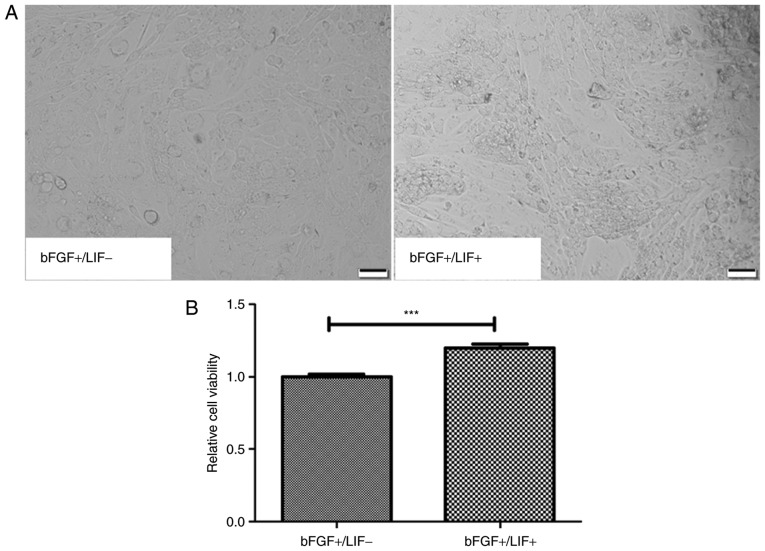
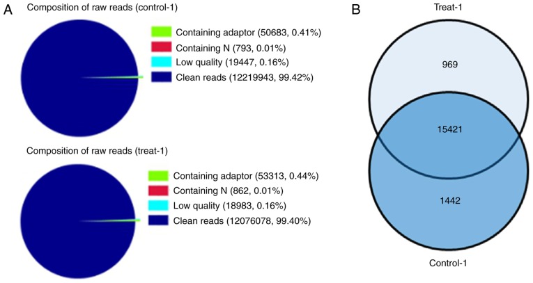
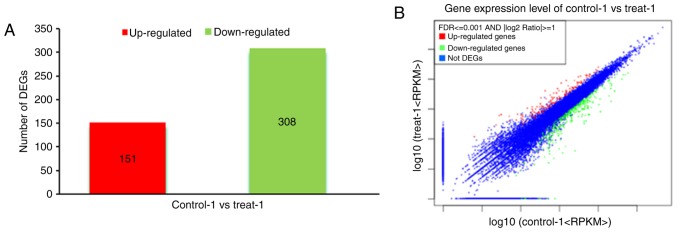
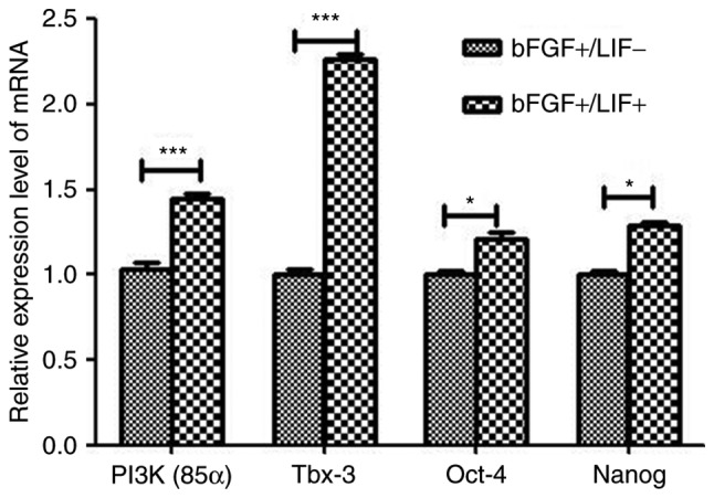
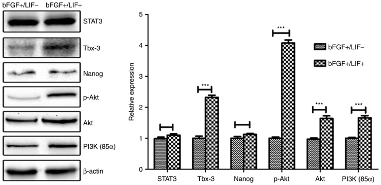
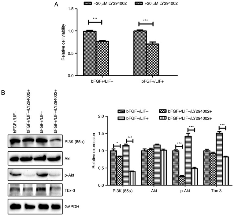
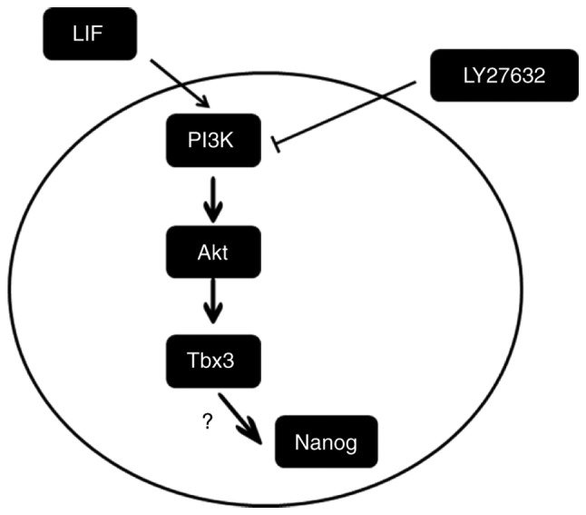
Leukemia inhibitory factor (LIF) is the most pleiotropic cytokine of the interleukin‑6 family, and is widely used to establish and maintain pluripotent stem cells, particularly mouse pluripotent stem cells. However, no reports have fully elucidated the application of LIF in marmoset induced pluripotent stem cell (iPSC) culture, particularly the underlying mechanisms. To demonstrate the feasibility of the application of LIF to marmoset iPSCs, the present study assessed these cells in the presence of LIF. Cell proliferation was measured using MTT assay, cell apoptosis was determined by flow cytometric analysis of fluorescein isothiocyanate Annexin V staining and the differentially expressed genes were analysed using Digital Gene Expression (DGE) analysis. The altered expression of pluripotency‑associated genes was confirmed by reverse transcription‑quantitative polymerase chain reaction and western blot analysis. Furthermore, following treatment with LY294002, cell proliferation was measured by MTT assay and protein levels were confirmed by western blot analysis. The results showed that LIF significantly promoted the number of proliferating cells, but had no effect on apoptosis. Digital Gene Expression analysis was used to examine the differentially expressed genes of marmoset iPSCs in the presence of LIF. The results showed that the pluripotency‑associated transcription factor‑encoding gene T‑box 3 (Tbx‑3) was activated by LIF. Notably, LIF increased the levels of phosphorylated (p‑)AKT and Tbx‑3 in the marmoset iPSCs. Furthermore, pretreatment with LY294002, an inhibitor of phosphoinositide 3‑kinase (PI3K), significantly impaired the LIF‑induced upregulation of p‑AKT and Tbx‑3 in the marmoset iPSCs, suggesting that the PI3K/Akt signaling pathway is involved in this regulation. Taken together, the results suggested that LIF is effective in maintaining marmoset iPSCs in cultures, which is associated with the activation of Tbx‑3 through regulation of the PI3K/Akt signaling pathway.


Figures

Figure 1
Figure 1
Effects of LIF on proliferation of marmoset iPSCs in culture. (A) Morphology and proliferation of marmoset iPSC clones cultured in the presence or absence of LIF for 24 h. The cloning efficiency of marmoset iPSCs was visibly increased in the presence of LIF (scale bar=50 µm). (B) Cell proliferation was measured using the MTT assay. The cell number in the control group cultured in the absent of LIF, was set as 1.0. ***P<0.001 (n=3). LIF, leukemia inhibitory factor; iPSCs, induced pluripotent stem cells; bFGF, basic fibroblast growth factor.
Figure 2
Figure 2
Effects of LIF on the apoptosis of marmoset iPSCs in culture. (A) Cell apoptosis was analyzed using flow cytometric analysis of FITC Annexin V staining. FITC Annexin V and PI-positive cells were analyzed by flow cytometry. (B) The majority of marmoset iPSCs were FITC Annexin V and PI negative. Apoptosis in the treatment group was 4.81±1.51% and in the control group was 4.25±1.01%. No significant difference in apoptosis was found. Q1, necrotic cells; Q2, late apoptosis; Q3, early apoptosis; Q4, viable cells; LIF, leukemia inhibitory factor; iPSCs, induced pluripotent stem cells; bFGF, basic fibroblast growth factor.
Figure 3
Figure 3
Differential expression of genes between the LIF-treated and control groups. (A) Sequencing quality assessment in each Digital Gene Expression library. Control-1, marmoset iPSCs cultured in the absence of LIF; Treat-1, marmoset iPSCs cultured in the presence of LIF. Different colors indicate the proportion distribution of reads. The data in parentheses indicate the number and percentage of corresponding tags among the total raw reads. Clean reads represents the raw sequence data without impurity, which is the basis for subsequent information analysis. (B) Venn diagram of the differentially expressed genes shared among the control and treatment groups. There were 15,421 genes shared between the two groups, and 969 and 1,442 genes were independently expressed by the treatment and control groups, respectively. iPSCs, induced pluripotent stem cells; LIF, leukemia inhibitory factor.
Figure 4
Figure 4
Volcano plot for the control and treated datasets. (A) Red indicates the upregulated genes in the LIF-treated group compared with the control group. Green indicates the downregulated genes in the LIF-treated group. (B) Results showing the number of upregulated and downregulated genes between the control and treated datasets. Blue indicates genes without expression differences between the two samples. LIF, leukemia inhibitory factor; DEGs, differentially expressed genes.
Figure 5
Figure 5
Altered expression of pluripotency-associated genes confirmed by reverse transcription-quantitative polymerase chain reaction analysis. The results showed the significant upregulation of Tbx-3 and PI3K(85α) (P<0.01). The mRNA level of the control group cultured in the absence of LIF was set as 1. *P<0.05 and ***P<0.001 (n=3). LIF, leukemia inhibitory factor; bFGF, basic fibroblast growth factor; Tbx-3, T-box 3; PI3K, phosphoinositide 3-kinase; Oct-4, octamer-binding protein 4.
Figure 6
Figure 6
Involvement of the PI3K/Akt signaling pathway in LIF-induced activation of Tbx-3 in marmoset induced pluripotent stem cells. The protein expression levels of STAT3, Tbx3, Nanog, p-Akt and PI3K(85α) in the presence or absence of LIF were analyzed by western blot analysis. Actin is shown as a loading control. ***P<0.001 (n=3). LIF, leukemia inhibitory factor; bFGF, basic fibroblast growth factor; STAT3, signal transducer and activator of transcription 3; Tbx-3, T-box 3; PI3K, phosphoinositide 3-kinase; p-, phosphorylated.
Figure 7
Figure 7
LY294002 suppresses cell viability. (A) Cell proliferation was measured using an MTT assay. LY294002, a PI3K inhibitor, reduced the LIF-stimulated proliferation of marmoset induced pluripotent stem cells. The number of cells in the control group cultured without LY294002 was set as 1.0. *P<0.05 and ***P<0.001 (n=3). (B) LIF increased the protein levels of PI3K(85α), p-AKT and TBX-3, and these effects were eradicated by the PI3K inhibitor (LY294002; 20 µM). GAPDH is shown as a loading control. LIF, leukemia inhibitory factor; bFGF, basic fibroblast growth factor; Tbx-3, T-box 3; PI3K, phosphoinositide 3-kinase; p-, phosphorylated.
Figure 8
Figure 8
Model of LIF-mediated expression levels of pluripotency-associated gene by activation of the PI3K/Akt signaling pathway. LIF, leukemia inhibitory factor; PI3K, phosphoinositide 3-kinase; Tbx-3, T-box 3.

Similar articles

Cited by

References

    1. De Vos J, Bouckenheimer J, Sansac C, Lemaître JM, Assou S. Human induced pluripotent stem cells: A disruptive innovation. Curr Res Transl Med. 2016;64:91–96. doi: 10.1016/j.retram.2016.04.001. - DOI - PubMed
    1. Wu Y, Mishra A, Qiu Z, Farnsworth S, Tardif SD, Hornsby PJ. Nonhuman primate induced pluripotent stem cells in regenerative medicine. Stem Cells Int. 2012;2012:767195. doi: 10.1155/2012/767195. - DOI - PMC - PubMed
    1. Thomson JA, Kalishman J, Golos TG, Durning M, Harris CP, Hearn JP. Pluripotent cell lines derived from common marmoset (Callithrix jacchus) blastocysts. Biol Reprod. 1996;55:254–259. doi: 10.1095/biolreprod55.2.254. - DOI - PubMed
    1. Sasaki E, Hanazawa K, Kurita R, Akatsuka A, Yoshizaki T, Ishii H, Tanioka Y, Ohnishi Y, Suemizu H, Sugawara A, et al. Establishment of novel embryonic stem cell lines derived from the common marmoset (Callithrix jacchus) Stem Cells. 2005;23:1304–1313. doi: 10.1634/stemcells.2004-0366. - DOI - PubMed
    1. Müller T, Fleischmann G, Eildermann K, Mätz-Rensing K, Horn PA, Sasaki E, Behr R. A novel embryonic stem cell line derived from the common marmoset monkey (Callithrix jacchus) exhibiting germ cell-like characteristics. Hum Reprod. 2009;24:1359–1372. doi: 10.1093/humrep/dep012. - DOI - PubMed

MeSH terms

Substances