

Expression and clinical significance of PD‑1 in hepatocellular carcinoma tissues detected by a novel mouse anti-human PD‑1 monoclonal antibody
- PMID: 29620156
- PMCID: PMC6929674
- DOI: 10.3892/ijo.2018.4358
Expression and clinical significance of PD‑1 in hepatocellular carcinoma tissues detected by a novel mouse anti-human PD‑1 monoclonal antibody
Abstract
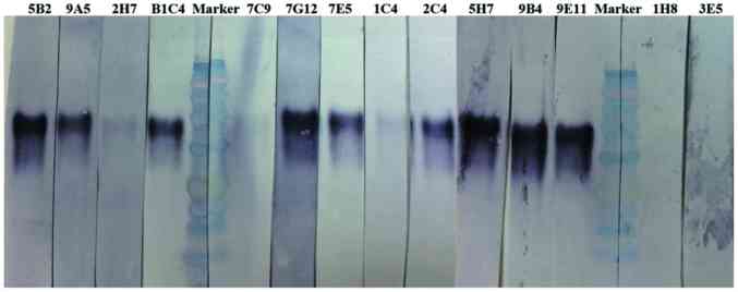
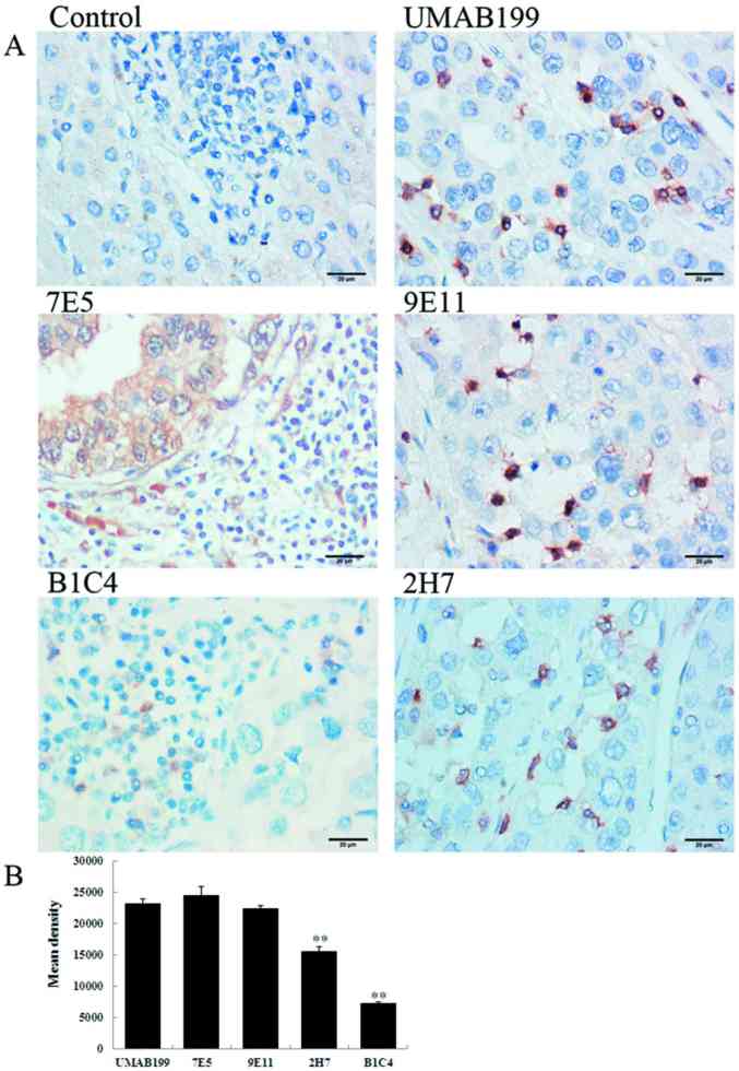
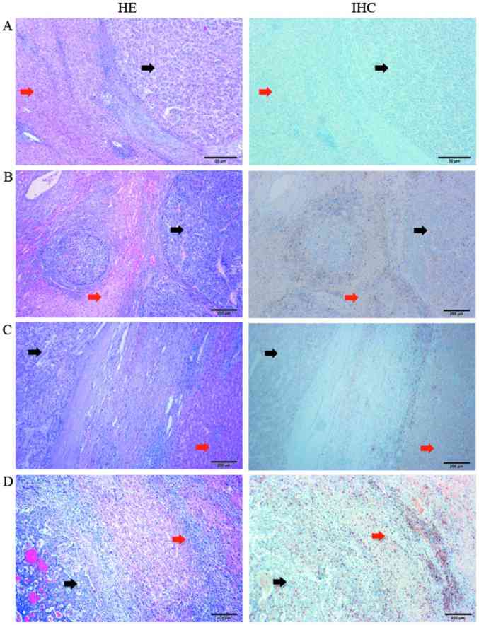
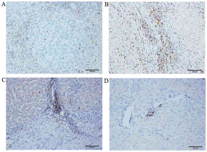
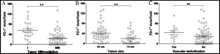
Hepatocellular carcinoma (HCC) is one of the most common malignancies and causes of death worldwide. Research investigating novel therapeutic strategies for the treatment of HCC is urgently required. Monoclonal antibodies (mAbs) that target the programmed cell death‑1 (PD‑1/PDCD1)/programmed death-ligand 1 (PD-L1) immune checkpoint have demonstrated substantial clinical benefit for a variety of solid tumors; however, these mAbs have not been well studied in HCC. In the present study, Sp2/0-Ag14 myeloma cells and spleen cells derived from BALB/c mice immunized with the recombinant human PD‑1/PDCD1 protein were fused for the production of novel antibodies. The 9E11 mAb, which exhibited the highest specificity for PD‑1 in HCC tissues in western blot and immunohistochemical staining analyses, was used to investigate the clinical significance of PD‑1 expression in HCC tissues from 77 cases, which were collected and examined histologically. Overexpression of PD‑1 was identified in peritumoral tissues, primarily in the liver portal region. Importantly, by analyzing the clinical data from 77 HCC patients, the expression of PD‑1 was observed to be significantly correlated with larger tumor size (>5 cm) and poorly differentiated tumors. In addition, PD‑1 expression was moderately correlated with venous thrombosis, but not correlated with patient sex or age, liver cirrhosis, hepatitis B, tumor, node and metastasis (TNM) stage or tumor location. The results of the present study suggest that high-level PD‑1 expression may be an important factor associated with the immune checkpoint pathway in HCC. The results suggest that PD‑1 serves an important role in tumor immune evasion and may be a valuable immunodiagnostic marker. In addition, PD‑1 may serve as a therapeutic target for patients presenting with poorly differentiated HCC, thus indicating the potential application of a PD‑1 inhibitor for the treatment of HCC patients.
Figures
References
-
- Benson DM, Jr, Bakan CE, Mishra A, Hofmeister CC, Efebera Y, Becknell B, Baiocchi RA, Zhang J, Yu J, Smith MK, et al. The PD-1/PD-L1 axis modulates the natural killer cell versus multiple myeloma effect: A therapeutic target for CT-011, a novel monoclonal anti-PD-1 antibody. Blood. 2010;116:2286–2294. doi: 10.1182/blood-2010-02-271874. - DOI - PMC - PubMed
-
- Tykodi SS, Brahmer JR, Hwu WJ, Chow LQ, Topalian SL, Hwu P, Odunsi K, Camacho LH, Kauh JS, Pitot HC, et al. PD-1/PD-L1 pathway as a target for cancer immunotherapy: Safety and clinical activity of BMS-936559, an anti-PD-L1 antibody, in patients with solid tumors. J Clin Oncol. 2012;30:2510.
-
- Horn L, Herbst RS, Spigel D, Gettinger SN, Gordon MS, Hollebecque, Kowanetz M. An analysis of the relationship of clinical activity to baseline EGFR status, PD-L1 expression and prior treatment history in patients with non-small cell lung cancer (NSCLC) following PD-L1 blockade with MPDL3280A (anti-PDL1) J Thorac Oncol. 2013;8:S364.
MeSH terms
Substances
LinkOut - more resources
Full Text Sources
Other Literature Sources
Medical
Research Materials
