. 2018 Jun;52(6):1863-1874.
doi: 10.3892/ijo.2018.4361. Epub 2018 Apr 5.

Increased level of cell-derived microparticles in the cyst fluids of odontogenic keratocysts

Affiliations

Increased level of cell-derived microparticles in the cyst fluids of odontogenic keratocysts

Qi-Wen Man et al. Int J Oncol. 2018 Jun.

Abstract

The aim of this study was to examine the level and basic characteristics of cell‑derived microparticles (MPs) in the cyst fluids of odontogenic keratocysts (OKCs). For this purpose, MPs from the cyst fluids (CFMPs) of OKCs were purified by a classic differential centrifugation method and characterized by a transmission electron microscope and fluorescence microscope. Flow cytometric analysis was used to determine the size, concentration and cellular origins of the CFMPs. Moreover, the expression level of receptor activator for nuclear factor‑κB ligand in the OKCs was evaluated by immunohistochemical staining and then analyzed for its correlation with the concentration of CFMPs by Spearman's rank correlation test. In addition, reverse transcription‑quantitative polymerase chain reaction (RT‑qPCR) and tartaric‑resistant acid phosphatase (TRAP) staining were performed to examine the osteoclastogenesis of mouse bone marrow‑derived macrophages (BMMs) in response to CFMPs. The results revealed that the levels of total CFMPs were significantly elevated in OKCs compared with dentigerous cysts (DCs) and radicular cysts (RCs). In addition, in vitro experiments further revealed that CFMPs derived from the OKCs of patients could be taken up by BMMs, leading to a significant increase in the mRNA expression levels of nuclear factor of activated T‑cells 1 (NFATc1) and TRAP. Moreover, TRAP‑positive multinucleated osteoclasts were successfully cultured in the presence of macrophage colony‑stimulating factor (M‑CSF) and CFMPs with BMMs. On the whole, our findings indicate that patients with OKCs have higher levels of CFMPs compared with patients with DCs and RCs, which may be associated with the bone resorption of OKCs.


Figures
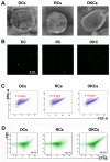
Figure 1
Figure 1
Identification of cyst fluid microparticles (CFMPs) derived from patients with dentigerous cysts (DCs), radicular cysts (RCs) and odontogenic keratocysts (OKCs). (A) Transmission electron microscopy (TEM) images of CFMPs purified from DCs, RCs and OKCs. (B) Fluorescence images showing the successful labeling of CFMPs by the fluorescent dye, carboxyfluorescein succinimidyl ester (CFSE). (C) The size distribution of CFMPs derived from DCs, RCs and OKCs was characterized by flow cytometry based on Nile Red fluorescent particles (red) with known diameters ranging from 0.7–0.9 µm. (D) Flow cytometry images showing that CFMPs derived from patients with DCs, RCs and OKCs could be largely stained with CFSE.
Figure 2
Figure 2
Cellular origin of cyst fluid microparticles (CFMPs). To determine the cellular origin, antibodies were used directly against CD31+/CD41+ (platelets), CD235a+/Annexin V+ (erythrocyte), CD144+/Annexin V+ (endothelium), CD45+/Annexin V+ (leukocyte) and EpCAM+/Annexin V+ (epithelium). Data are expressed and sorted by the number of microparticles per microlitre.
Figure 3
Figure 3
Quantification of the subtypes of cyst fluid microparticles (CFMPs). (A–E) Quantitative analysis of the levels of CD31+/CD41+ (platelets) CFMPs, CD235a+/Annexin V+ (erythrocytes) CFMPs, CD144+/Annexin V+ (endothelium) CFMPs, CD45+/Annexin V+ (leukocytes) CFMPs and EpCAM+/Annexin V+ (epithelium) CFMPs in patients with dentigerous cysts (DCs), radicular cysts (RCs) and odontogenic keratocysts (OKCs). Data are expressed as the means ± SEM. *P<0.05 vs. the control group.
Figure 4
Figure 4
Quantification of the level of cyst fluid microparticles (CFMPs) and the correlation between the level of CFMPs and the clinical characteristics of patients with dentigerous cysts (DCs), radicular cysts (RCs) and odontogenic keratocysts (OKCs). (A) Representative flow cytometry dot-plots showing gated CFMPs (green dots) and the fluorescent beads for counting (red dots). (B) Quantitative analysis of the level of CFMPs derived from patients with DCs, RCs and OKCs. (C) Representative immunohistochemical staining of receptor activator for nuclear factor-κB ligand (RANKL) in OKCs with different level of CFMPs. (D) Quantitative analysis of staining intensity of RANKL in OKCs with different levels of CFMPs. (E) Spearman's rank correlation test was performed to determine the correlation between the level of CFMPs and the mean density of RANKL in OKCs. The data are expressed as the means ± SEM. *P<0.05 vs. the control group; N. S, not significant.
Figure 5
Figure 5
Effect of cyst fluid microparticles (CFMPs) isolated from two patients with odontogenic keratocysts (OKCs) on human immortalized oral epithelial cells (HIOECs). (A) Uptake assays for CFSE-labeled CFMPs (green) were performed by using HIOECs (red) as the recipient cells. (B) DNA electrophoresis indicated that CFMPs from OKCs expressed receptor activator for nuclear factor-κB ligand (RANKL) mRNA. (C) RANKL expression was examined by western blot analysis in CFMPs co-cultured with HIOECs. The CFMPs were selected from two patients with OKCs.
Figure 6
Figure 6
Cells were treated with or without CFMPs (5 or 10 μg/ml) in a 96-well plate for 24 or 48 h. Cell viability was measured by MTT assay. Results were expressed as the percentage (%) viability of cells compared to the control (means ± SEM, n=5). P-values <0.05 were considered to indicate statistically significant differences. No significant differences were observed in this analysis. The results shown here as a representative of 3 independent experiments. HIOECs, human immortalized oral epithelial cells; BMMs, bone marrow-derived macrophages.
Figure 7
Figure 7
Receptor activator for nuclear factor-κB ligand (RANKL) expression in cyst fluid microparticles (CFMPs) of jaw cysts. (A) Statistic analysis showing the RANKL7Annexin V+ microparticles in patients with dentigerous cysts (DCs), radicular cysts (RCs) and odontogenic keratocysts (OKCs). (B) The protein level of RANKL was examined by western blot analysis of the CFMPs from 4 patients with OKCs.
Figure 8
Figure 8
Effect of cyst fluid microparticles (CFMPs) isolated from 2 patients with odontogenic keratocysts (OKCs) on the osteoclastogenesis of bone marrow-derived macrophages (BMMs). (A) Uptake assays for CFSE-labeled CFMPs (green) were performed by using BMMs (labeled with CellMask) as the recipient cells. (B) Tartaric-resistant acid phosphatase (TRAP) staining showed that multinucleated osteoclasts were observed in the presence of macrophage colony-stimulating factor (M-CSF) (20 ng/ml) and CFMPs (5 or 10 µg/ml). The anti-receptor activator for nuclear factor-κB ligand (RANKL) monoclonal antibody was used to neutralize the RANKL protein. The soluble RANKL (sRANKL; 50 ng/ml)-treated group was selected as a positive control. (C) Quantification analysis of the results from TRAP staining. (D and E) The mRNA levels of the nuclear factor of activated T-cells 1 (NFATc1) and TRAP were evaluated by RT-qPCR in BMMs co-cultured with various concentrations of CFMPs. The M-CSF (20 ng/ml)- and sRANKL (50 ng/ml)-treated group was used as a positive control. Data are shown as the means ± SEM. *P<0.05, **P<0.01 and ***P<0.001 vs. the control group. #P<0.05 and ##P<0.01 vs. the CFMP (5 µg/ml)-treated group from the same patient. ^^P<0.05 and ^^^P< 0.01 vs. the CFMP (10 µg/ml)-treated group from the same patient.

Similar articles

Cited by

References

    1. Browne RM. The pathogenesis of odontogenic cysts: A review. J Oral Pathol. 1975;4:31–46. doi: 10.1111/j.1600-0714.1975.tb01738.x. - DOI - PubMed
    1. Kramer IR, Pindborg JJ, Shear M. The WHO histological typing of odontogenic tumours. A commentary on the second edition. Cancer. 1992;70:2988–2994. doi: 10.1002/1097-0142(19921215)70:12<2988::AID-CNCR2820701242>3.0.CO;2-V. - DOI - PubMed
    1. Avelar RL, Antunes AA, Carvalho RW, Bezerra PG, Oliveira Neto PJ, Andrade ES. Odontogenic cysts: A clinicopathological study of 507 cases. J Oral Sci. 2009;51:581–586. doi: 10.2334/josnusd.51.581. - DOI - PubMed
    1. Lin HP, Wang YP, Chen HM, Cheng SJ, Sun A, Chiang CP. A clinicopathological study of 338 dentigerous cysts. J Oral Pathol Med. 2013;42:462–467. doi: 10.1111/jop.12042. - DOI - PubMed
    1. Thompson L. World Health Organization classification of tumours: Pathology and genetics of head and neck tumours. Ear Nose Throat J. 2006;85:74. - PubMed

MeSH terms