. 2018 Jul;42(1):115-122.
doi: 10.3892/ijmm.2018.3598. Epub 2018 Mar 29.

Tripterygium glycoside protects against puromycin amino nucleoside‑induced podocyte injury by upregulating autophagy

Affiliations

Tripterygium glycoside protects against puromycin amino nucleoside‑induced podocyte injury by upregulating autophagy

Jianguang Gong et al. Int J Mol Med. 2018 Jul.

Abstract

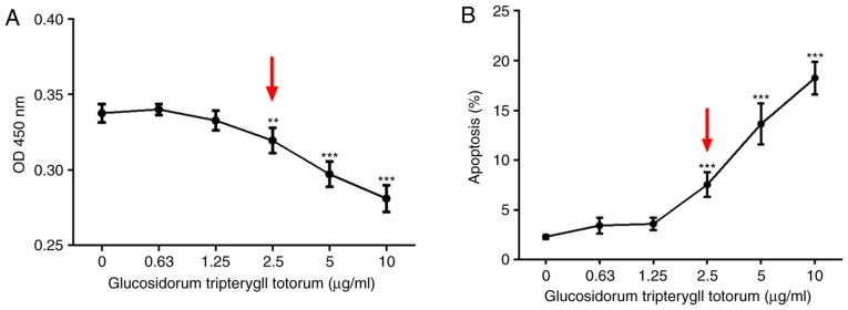
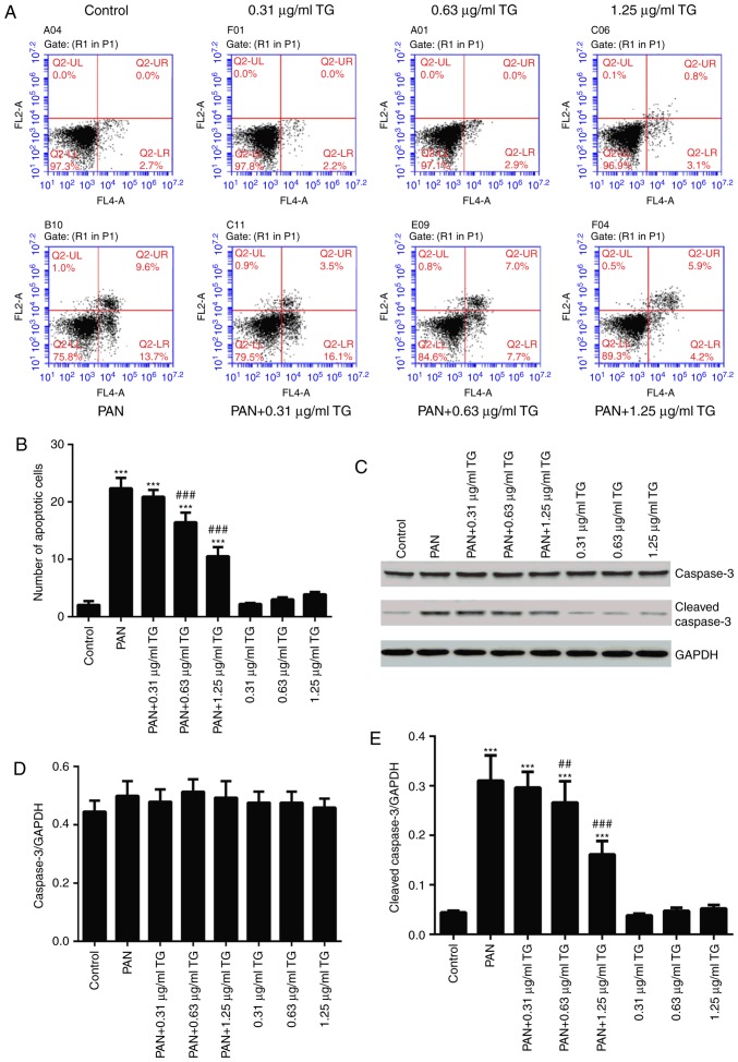
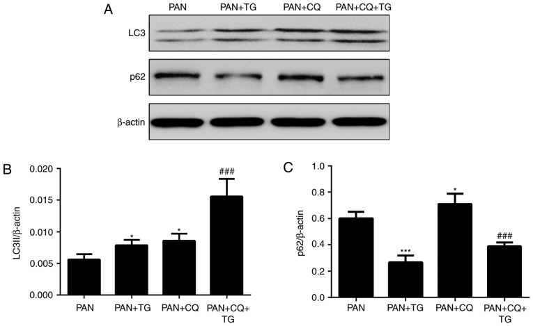
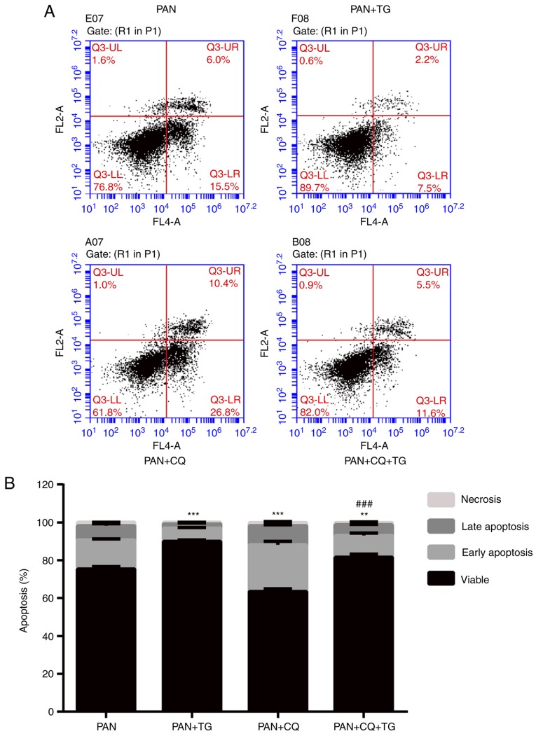
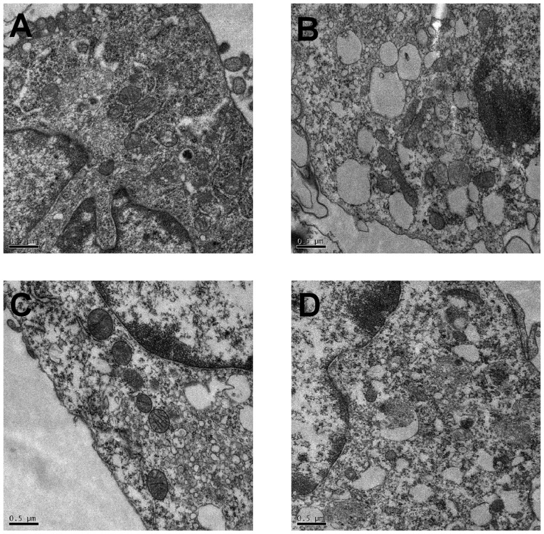
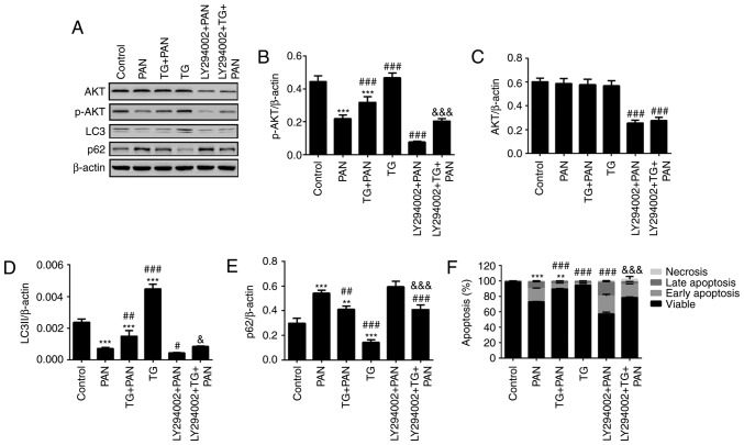
Tripterygium glycoside (TG), an active ingredient of the widely used Chinese herb Tripterygium wilfordii Hook F, has immunosuppressive and anti‑inflammatory effects. Previous studies have indicated that TG is a potentially effective therapeutic option to treat nephrotic syndrome. The mechanism underlying the therapeutic effect of TG, including its effect on autophagy and apoptosis in podocyte injury, remains to be fully elucidated. The present study aimed to assess the protective effect of TG on podocytes via its potential role in the activation of autophagic and phosphatidylinositol 3‑kinase (PI3K) pathways. Using flow cytometry, western blot analysis, cell counting kit‑8 assays and transmission electron microscopy analysis, the effects of TG on puromycin aminonucleoside (PAN)‑induced podocyte injury were investigated. Chloroquine (CQ), an inhibitor of autophagy, was used to assess the importance of autophagy in the protective effect of TG. In addition, LY294002, an inhibitor of class III PI3K, was used to identify which signaling pathways TG is involved in. PAN caused marked apoptosis of podocytes, which was significantly antagonized by TG. The expression of microtubule‑associated protein 1A/1B‑light chain 3 and the appearance of autophagosomes increased significantly following TG treatment, whereas the expression levels of p62 and cleaved caspase-3 were markedly decreased. Podocyte apoptosis decreased significantly when the podocytes were treated with TG compared with the levels of apoptosis in the PAN‑ and PAN+CQ‑treated groups. The expression of phosphorylated AKT was increased significantly in the TG‑treated groups, and the effects of TG on the podocytes were significantly inhibited by LY294002. In conclusion, TG protected podocytes from PAN‑induced injury, and the effects were attributable to the activation of autophagy, mainly via a PI3K‑dependent pathway.


Figures

Figure 1
Figure 1
Cytotoxic effect of TG on cultured podocytes. (A) Cell counting kit-8 assay results of MPCs following treatment with different TG concentrations. At a TG concentration of 2.5 µg/ml, cell viability was significantly decreased (P<0.01; red arrow). (B) Apoptotic rates of MPCs following treatment with different TG concentrations, as detected by the Annexin V/propidium iodide assay. The percentage of apoptotic cells increased significantly at a TG concentration of 2.5 µg/ml (P<0.001; red arrow). Data are presented as the mean ± standard deviation (n=3). **P<0.01 and ***P<0.001 vs. 0 µg/ml group. MPCs, mouse podocyte cells; TG, tripterygium glycoside.
Figure 2
Figure 2
Protective effect of TG on the apoptosis of PAN-treated podocytes. (A) Flow cytometric dot plots of Annexin V and propidium iodide staining. (B) Bar graph showing the percentage of cells undergoing early apoptosis. (C) Representative western blots of cleaved-caspase-3 and caspase-3 in each group. (D) Bar graph showing the relative expression of caspase-3. No significant differences between the groups were observed. (E) Bar graph showing the relative expression of cleaved-caspase-3. The expression of cleaved-caspase-3 was markedly increased in the PAN group, compared with that in the control group, and was decreased significantly in the PAN+0.63 µg/ml TG group and PAN+1.25 µg/ml TG group, compared with that in the PAN group. Data are presented as the mean ± standard deviation (n=3). ***P<0.001 vs. control group; ##P<0.01 and ###P<0.001 vs. PAN group. TG, tripterygium glycoside; PAN, puromycin aminonucleoside.
Figure 3
Figure 3
Effects of TG on the autophagy of podocytes. The levels of autophagic markers were detected by western blot analysis. (A) Representative bands of LC3 and p62 in the individual groups. Bar graphs show the relative protein expression of LC3II and p62. (B) TG and CQ promoted the expression of LC3II in podocytes. (C) Expression of p62 was markedly decreased in the PAN+TG group and PAN+CQ+TG group, compared with that in the PAN group and the PAN+CQ group, respectively. Data are presented as the mean ± standard deviation (n=3). *P<0.05 and ***P<0.001 vs. PAN group; ###P<0.001 vs. PAN+CQ group. TG, tripterygium glycoside; PAN, puromycin aminonucleoside; CQ, chloroquine.
Figure 4
Figure 4
TG protects podocytes from apoptosis through the upregulation of autophagy. (A) Flow cytometric dot plots of Annexin V and propidium iodide staining. (B) Bar graph showing the rates of apoptosis of podocytes in the individual groups. The podocyte apoptotic rate was decreased in the PAN+TG group and the PAN+CQ+TG group, compared with the rate in the PAN group and PAN+CQ group, respectively. Data are presented as the mean ± standard deviation (n=3). *P<0.05, **P<0.01 and ***P<0.001 vs. PAN group; #P<0.05, ##P<0.01 and ###P<0.001 vs. PAN+CQ group. TG, tripterygium glycoside; PAN, puromycin aminonucleoside; CQ, chloroquine.
Figure 5
Figure 5
Autophagosomes detected by transmission electron microscopy in the podocytes of individual groups. Autophagosomes were detected by transmission electron microscopy (magnification, ×20,000). (A) Numerous autophagosomes were observed in the cytoplasm of podocytes in the control group. (B) Compared with the control group, the number of autophagosomes was markedly lower in the PAN-treated podocytes. (C) Compared with the control group, the number of autophagosomes was markedly increased following TG treatment. (D) Compared with the PAN group, the number of autophagosomes was increased following TG treatment. TG, tripterygium glycoside; PAN, puromycin aminonucleoside.
Figure 6
Figure 6
TG promotes the activation of autophagy by PI3K signaling during podocyte injury. (A) Representative bands of AKT, p-AKT, LC3 and p62 proteins in the individual groups. Quantitative evaluation of the levels of (B) p-AKT, (C) AKT, (D) LC3 and (E) p62 in the individual groups. The expression of p-AKT was markedly inhibited by LY294002. The expression of p-AKT was markedly increased in the TG+PAN and LY294002+TG+PAN groups, compared with the expression in the PAN group and LY294002+PAN group, respectively. TG promoted the expression of LC3II and decreased the expression of p62. (F) Apoptotic rates in the groups. Data are presented as the mean ± standard deviation (n=3). **P<0.01 and ***P<0.001 vs. control group; #P<0.05, ##P<0.01 and ###P<0.001 vs. PAN group; &P<0.05 and &&&P<0.001 vs. LY294002+PAN group. TG, tripterygium glycoside; PAN, puromycin aminonucleoside; PI3K, phosphatidylinositol 3-kinase; p-, phosphorylated; LC3, microtubule-associated protein 1A/1B-light chain 3.

References

    1. Cara-Fuentes G, Clapp WL, Johnson RJ, Garin EH. Pathogenesis of proteinuria in idiopathic minimal change disease: Molecular mechanisms. Pediatr Nephrol. 2016;31:2179–2189. doi: 10.1007/s00467-016-3379-4. - DOI - PubMed
    1. Mallipattu SK, He JC. The podocyte as a direct target for treatment of glomerular disease. Am J Physiol Renal Physiol. 2016;311:F46–F51. doi: 10.1152/ajprenal.00184.2016. - DOI - PMC - PubMed
    1. Nagata M. Podocyte injury and its consequences. Kidney Int. 2016;89:1221–1230. doi: 10.1016/j.kint.2016.01.012. - DOI - PubMed
    1. Xu X, Li QJ, Xia S, Wang MM, Ji W. Tripterygium glycosides for treating late-onset rheumatoid arthritis: A systematic review and meta-analysis. Altern Ther Health Med. 2016;22:32–39. - PubMed
    1. Chen Y, Gong Z, Chen X, Tang L, Zhao X, Yuan Q, Cai G. Tripterygium wilfordii Hook F (a traditional Chinese medicine) for primary nephrotic syndrome. Cochrane Database Syst Rev. 2013;8:CD008568. - PubMed

MeSH terms