. 2018 Jun;52(6):1899-1911.
doi: 10.3892/ijo.2018.4343. Epub 2018 Mar 29.

Metformin inhibits ovarian cancer via decreasing H3K27 trimethylation

Affiliations

Metformin inhibits ovarian cancer via decreasing H3K27 trimethylation

Guiju Tang et al. Int J Oncol. 2018 Jun.

Abstract

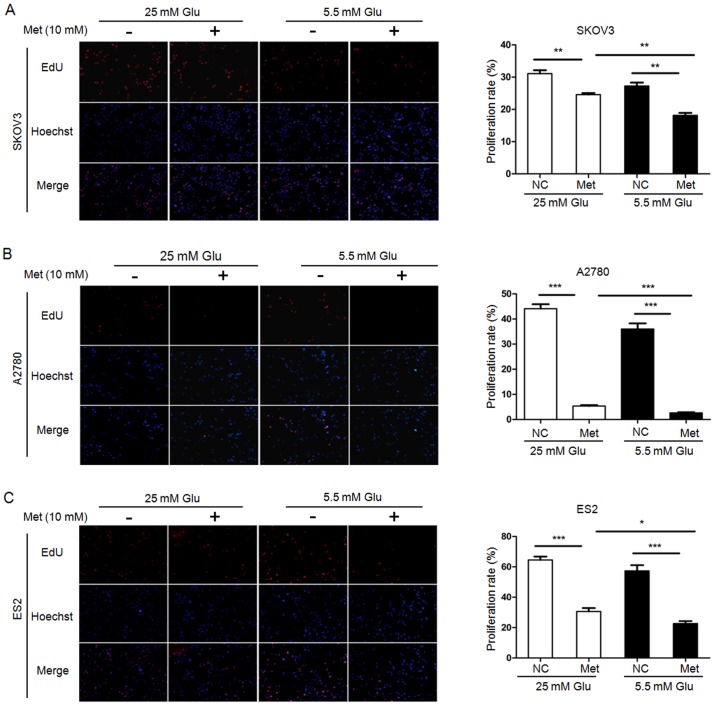
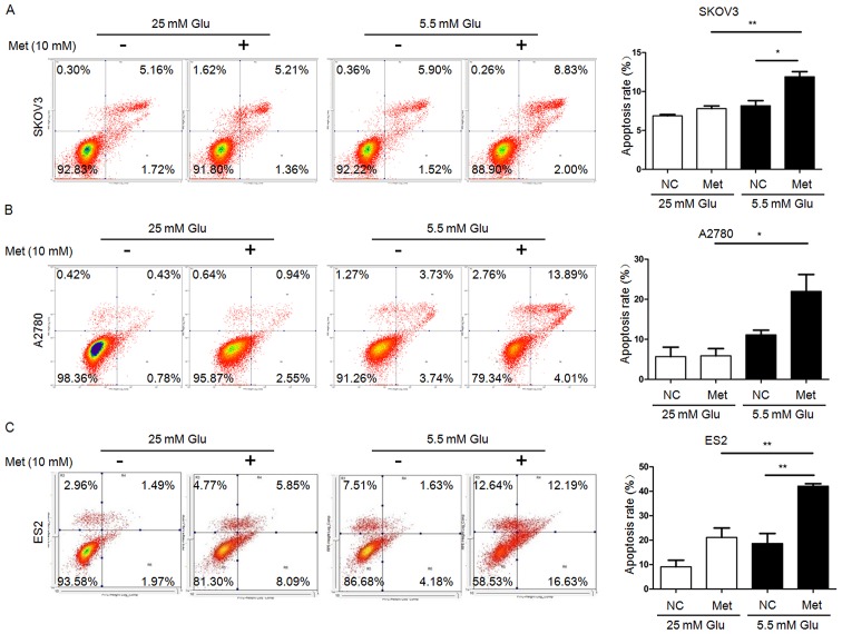
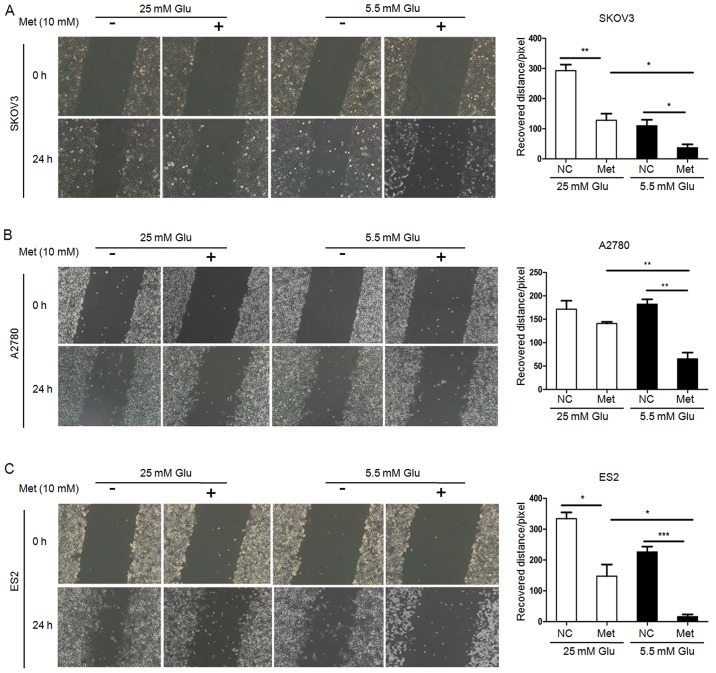
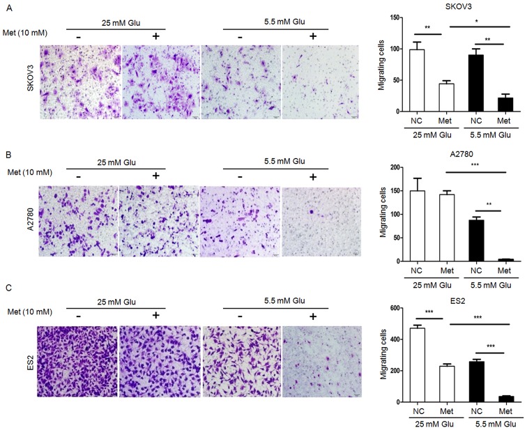
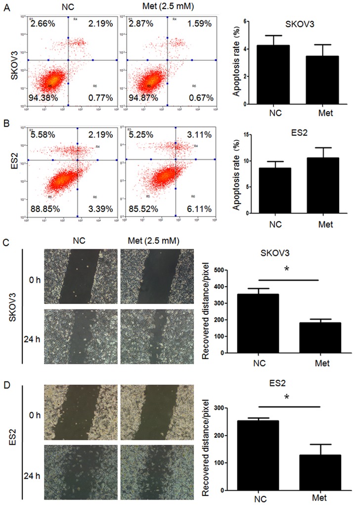
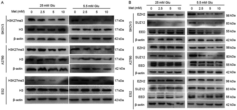
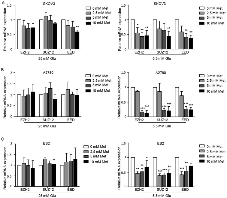
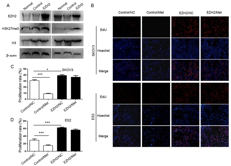
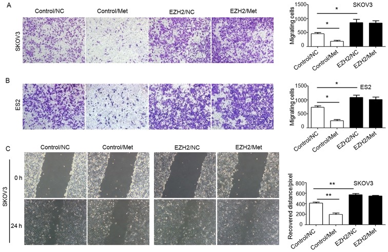
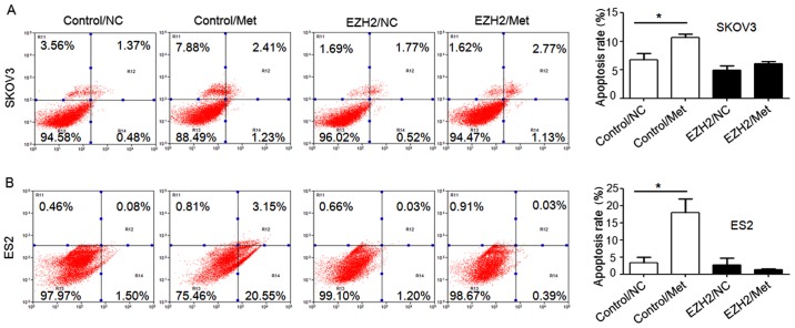
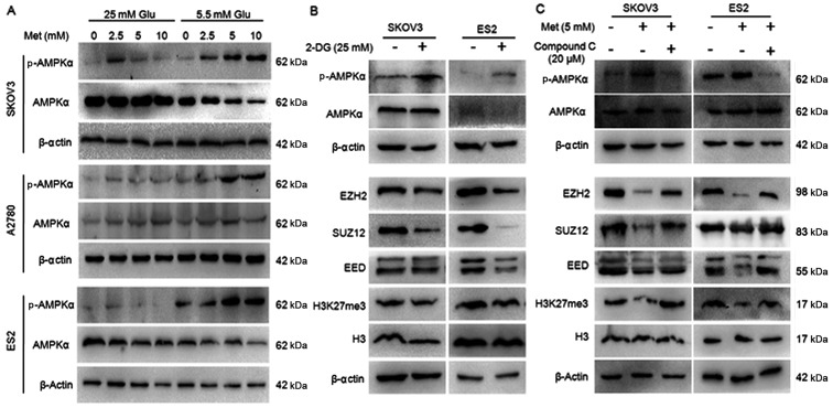
Metformin has been used for the treatment of type II diabetes mellitus for decades. Recently, used of metformin in the therapy of diverse human cancer types has received widespread attention, while the underlying mechanisms have been not fully elucidated. In the current study, 5-ethynyl-20-deoxyuridine assay to detect cell proliferation, flow cytometry to detect apoptosis, scratch wound healing and Transwell migration assay to detect cell migration capacity. The current study reported that metformin inhibited cell proliferation and migration, and promoted apoptosis in ovarian cancer cells, particularly under normoglycemic conditions in vitro. Metformin treatment significantly promoted the phosphorylation of AMP-activated protein kinase (AMPK), and reduced histone H3 lysine 27 trimethylation (H3K27me3) and polycomb repressor complex 2 (PRC2) levels. Additionally, overexpression of EZH2 to increase H3K27me3 abrogated the effect of metformin on the cell proliferation, migration and apoptosis in SKOV3 and ES2 cells. Similar to metformin, another AMPK agonist, 2-deoxy-D-glucose, reduced the H3K27me3 level and PRC2 expression. In cells pretreated with Compound C, an AMPK inhibitor, metformin was not able to induce AMPK phosphorylation or reduce H3K27me3. Metformin-mediated AMPK activation and H3K27me3 inhibition were more robust in cells exposed to low glucose (5.5 mM) compared with those exposed to high glucose (25 mM). These findings implicate H3K27me3 repression mediated by AMPK phosphorylation in the antitumor effect of metformin in ovarian cancer, indicating that metformin alters epigenetic modifications by targeting PRC2 and supports the use of metformin in treatment of patients with epithelial ovarian cancer without diabetes.


Figures

Figure 1
Figure 1
Metformin inhibits the proliferation of ovarian cancer cells. EdU assays demonstrate that metformin reduces proliferation of (A) SKOV3, (B) A2780 and (C) ES2 cells. Representative fluorescence microscopy images of EdU incorporation and Hoechst 33342 staining of nuclei (magnification, ×100). The proliferation rates of SKOV3, A2780, and ES2 cells were calculated. Data are presented as the mean ± standard deviation. *P<0.05; **P<0.01; ***P<0.001. Met, metformin; Glu, glucose; EdU, 5-ethynyl-20-deoxyuridine; NC, negative control.
Figure 2
Figure 2
Metformin promotes the apoptosis of ovarian cancer cells. Percentage of apoptotic (A) SKOV3, (B) A2780 and (C) ES2 cells were detected by flow cytometry analysis using Annexin V-fluorescein isothiocyanate/PI staining. Apoptotic cells includes Annexin V(-)/PI(+), Annexin V(+)/PI(−) and Annexin V(+)/PI(+) cells. Data are presented the mean ± standard deviation. *P<0.05; **P<0.01. PI, propidium iodide; Met, metformin; Glu, glucose; NC, negative control.
Figure 3
Figure 3
Metformin inhibits migration of ovarian cancer cells. Migration of (A) SKOV3, (B) A2780 and (C) ES2 cells were measured by scratch wound healing assay. Representative bright-field images (magnification, ×100) and quantification of recovered distance at 24 h after 10 mM metformin treatment. Data are presented the mean ± standard deviation. *P<0.05; **P<0.01; ***P<0.001. Met, metformin; Glu, glucose; NC, negative control.
Figure 4
Figure 4
Metformin inhibits migration of ovarian cancer cells. Migration of (A) SKOV3, (B) A2780 and (C) ES2 cells were measured by Transwell migration assay. Representative bright-field images of the cell migration (magnification, ×100) and quantification of migrated cells at 24 h after 10 mM metformin treatment are shown. Data are presented the mean ± standard deviation. *P<0.05; **P<0.01;***P<0.001. Met, metformin; Glu, glucose; NC, negative control.
Figure 5
Figure 5
Metformin (2.5 mM) has no significant effect on apoptosis of ovarian cancer cells, however migration is inhibited. Percentage of apoptotic (A) SKOV3 and (B) ES2 cells were detected by flow cytometry analysis using Annexin V-fluorescein isothiocyanate/PI staining. Apoptotic cells include Annexin V(−)/PI(+), Annexin V(+)/PI(−) and Annexin V(+)/PI(+) cells. Migration of (C) SKOV3 and (D) ES2 were measured by scratch wound healing assay. Representative bright-field images (magnification, ×100) and quantification of recovered distance at 24 h after 2.5 mM metformin treatment. Data are presented the mean ± standard deviation. *P<0.05. PI, propidium iodide; Met, metformin; Glu, glucose; NC, negative control.
Figure 6
Figure 6
Metformin decreases H3K27me3 and expression of polycomb repressor complex 2 in ovarian cancer cells. The protein expression of (A) H3K27me3 and (B) EZH2, SUZ12 and EED in A2780, SKOV3 and ES2 cells were assayed by western blot analysis. Met, metformin; Glu, glucose; H3K27me3, histone H3 lysine 27 trimethylated; H3, histone H3; EZH2, histone-lysine N-methyltransferase EZH2; SUZ12, polycomb protein SUZ12; EED, polycomb protein EED.
Figure 7
Figure 7
Metformin decreases PRC2 of ovarian cancer cells. mRNA expression of EZH2, SUZ12 and EED in (A) SKOV3, (B) A2780 and (C) ES2 cells determined by reverse transcription-quantitative polymerase chain reaction analysis. Data are presented the mean ± standard deviation. *P<0.05; **P<0.01; ***P<0.001. Met, metformin; Glu, glucose; EZH2, histone-lysine N-methyltransferase EZH2; SUZ12, polycomb protein SUZ12; EED, polycomb protein EED.
Figure 8
Figure 8
Overexpression of EZH2suppresses the inhibitory effect of metformin on proliferation of ovarian cancer cells. (A) EZH2 DNA lentivirus upregulated EZH2 andH3K27me3 protein expression. The protein expression of EZH2 and the level of H3K27me3 in SKOV3 and ES2 cells were assayed by western blot analysis. (B) EdU assays evaluated the proliferation of ovarian cancer cells. Representative fluorescence microscopy images of EdU incorporation and Hoechst 33342 staining of nuclei (magnification, ×100). The proliferation rates of (C) SKOV3 and (D) ES2 cells. Data are presented the mean ± standard deviation. *P<0.05; ***P<0.001. EZH2, histone-lysine N-methyltransferase EZH2; H3K27me3, histone H3 lysine 27 trimethylated; H3, histone H3; NC, negative control; Met, metformin; EdU, 5-ethynyl-20-deoxyuridine.
Figure 9
Figure 9
Overexpression of EZH2 suppresses the inhibitory effect of metformin on migration of ovarian cancer cells. Migration of (A) SKOV3 and (B) ES2 cells were measured by Transwell migration assay (crystal violet staining), and (C) a scratch wound healing assay was performed using SKOV3 cells. Representative bright-field images of the cell migration (magnification, ×100) and quantification of migrated cells or recovered distance at 24 h after 10 mM metformin treatment are shown. Data are presented the mean ± standard deviation. *P<0.05; **P<0.01. NC, negative control; Met, metformin; EZH2, histone-lysine N-methyltransferase EZH2.
Figure 10
Figure 10
Overexpression of EZH2 suppresses the effect of metformin on apoptosis of ovarian cancer cells. Percentage of apoptotic (A) SKOV3 and (B) ES2 cells were detected by flow cytometry analysis using Annexin V-PE/7-AAD staining. Apoptotic cells include Annexin V(−)/7-AAD(+), Annexin V(+)/7-AAD(−) and Annexin V(+)/7-AAD(+) cells. Data are presented the mean ± standard deviation. *P<0.05. PE, phycoerythrin; 7-AAD, 7-aminoactinomycin D; NC, negative control; Met, metformin; EZH2, histone-lysine N-methyltransferase EZH2.
Figure 11
Figure 11
Metformin inhibits H3K27me3 through the AMPK pathway. (A) Metformin activates AMPK of ovarian cancer cells in normoglycemic condition. (B) 2-DG activates AMPK and suppresses H3K27me3, PRC2 of ovarian cancer cells in normoglycemic condition. (C) Compound C inhibits the effect of metformin on PRC2 and H3K27me3 when ovarian cancer cells were cultured in normoglycemic condition. The protein expression of p-AMPKα, AMPKα, H3K27me3, EZH2, SUZ12 and EED in A2780, SKOV3 and ES2 cells were assayed by western blot analysis. The expression was normalized to β-actin. Met, metformin; Glu, glucose; p-AMPKα, phospho-AMPKα; AMPKα, AMP-activated protein kinase; 2-DG, 2-deoxy-D-glucose; EZH2, histone-lysine N-methyltransferase EZH2;SUZ12, polycomb protein SUZ12; EED, polycomb protein EED; 2-DG, dorsomorphin 2HCl; H3K27me3, histone H3 lysine 27 trimethylation.

Similar articles

Cited by

References

    1. Siegel RL, Miller KD, Jemal A. Cancer statistics, 2016. CA Cancer J Clin. 2016;66:7–30. doi: 10.3322/caac.21332. - DOI - PubMed
    1. Davis A, Tinker AV, Friedlander M. 'Platinum resistant' ovarian cancer: What is it, who to treat and how to measure benefit? Gynecol Oncol. 2014;133:624–631. doi: 10.1016/j.ygyno.2014.02.038. - DOI - PubMed
    1. Coyle C, Cafferty FH, Vale C, Langley RE. Metformin as an adjuvant treatment for cancer: A systematic review and meta-analysis. Ann Oncol. 2016;27:2184–2195. doi: 10.1093/annonc/mdw410. - DOI - PMC - PubMed
    1. Dowling RJ, Niraula S, Chang MC, Done SJ, Ennis M, McCready DR, Leong WL, Escallon JM, Reedijk M, Goodwin PJ, et al. Changes in insulin receptor signaling underlie neoadjuvant metformin administration in breast cancer: A prospective window of opportunity neoadjuvant study. Breast Cancer Res. 2015;17:32. doi: 10.1186/s13058-015-0540-0. - DOI - PMC - PubMed
    1. Rattan R, Graham RP, Maguire JL, Giri S, Shridhar V. Metformin suppresses ovarian cancer growth and metastasis with enhancement of cisplatin cytotoxicity in vivo. Neoplasia. 2011;13:483–491. doi: 10.1593/neo.11148. - DOI - PMC - PubMed