. 2018 Jun;39(6):2482-2498.
doi: 10.3892/or.2018.6332. Epub 2018 Mar 23.

Translational control of the undifferentiated phenotype in ER‑positive breast tumor cells: Cytoplasmic localization of ERα and impact of IRES inhibition

Affiliations

Translational control of the undifferentiated phenotype in ER‑positive breast tumor cells: Cytoplasmic localization of ERα and impact of IRES inhibition

Christos Vaklavas et al. Oncol Rep. 2018 Jun.

Abstract

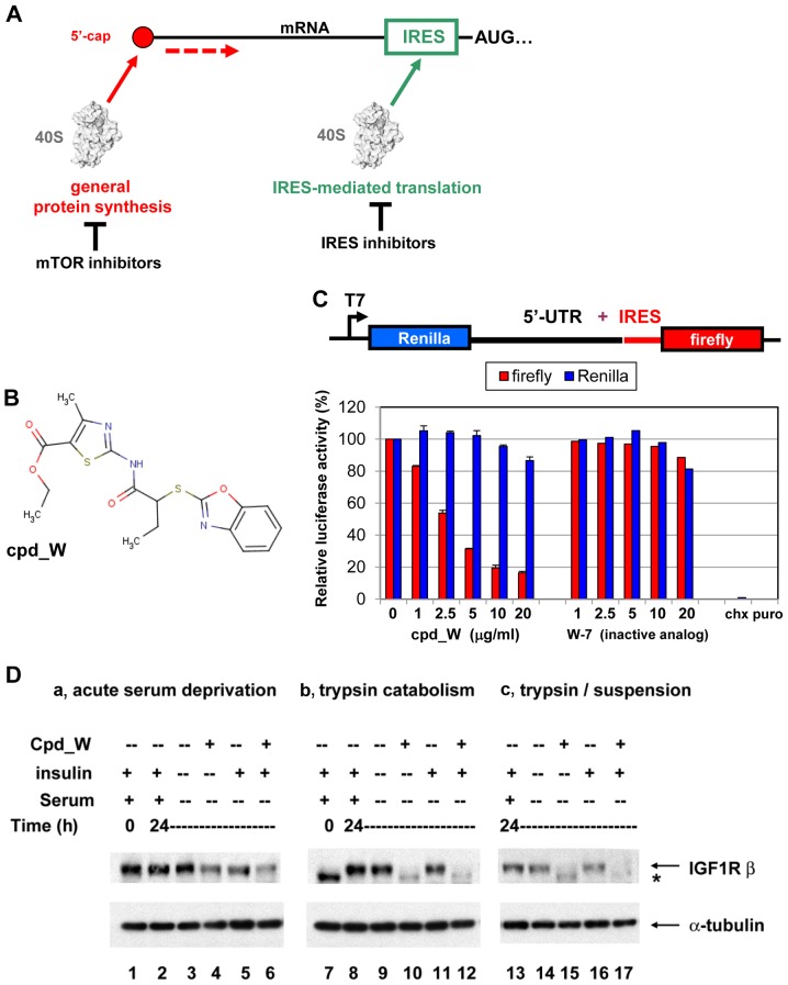
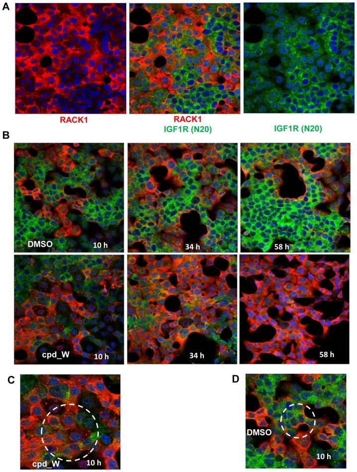
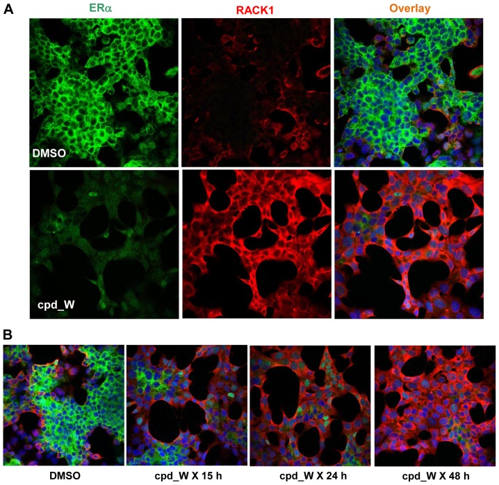
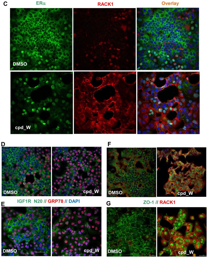
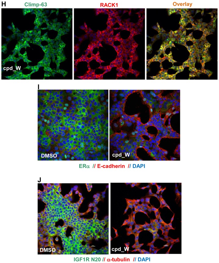
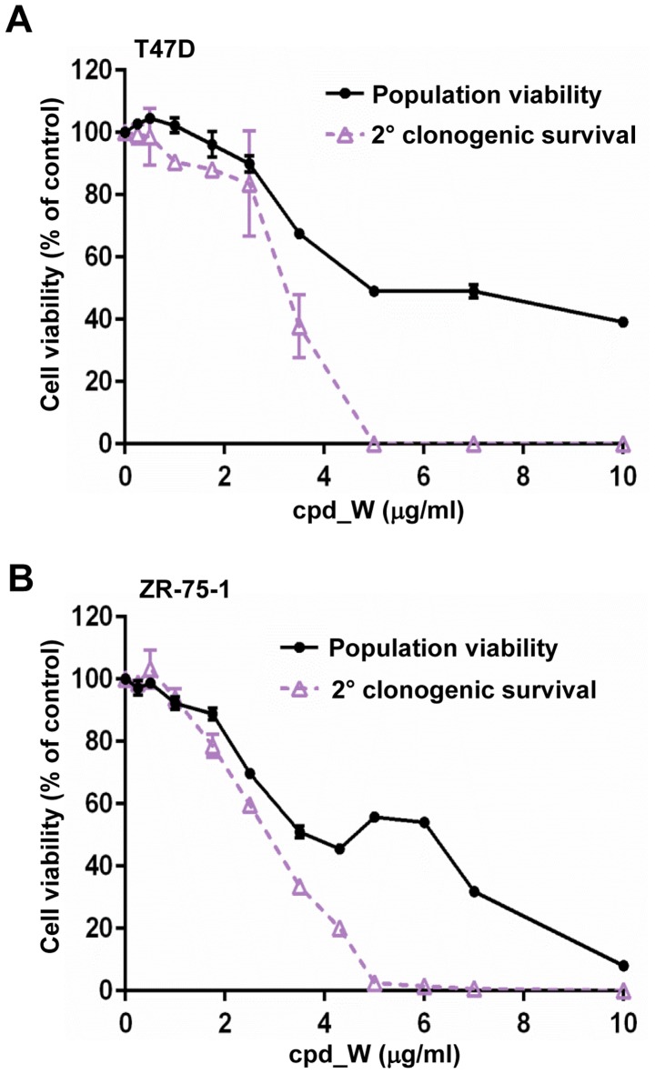
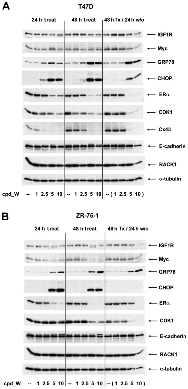
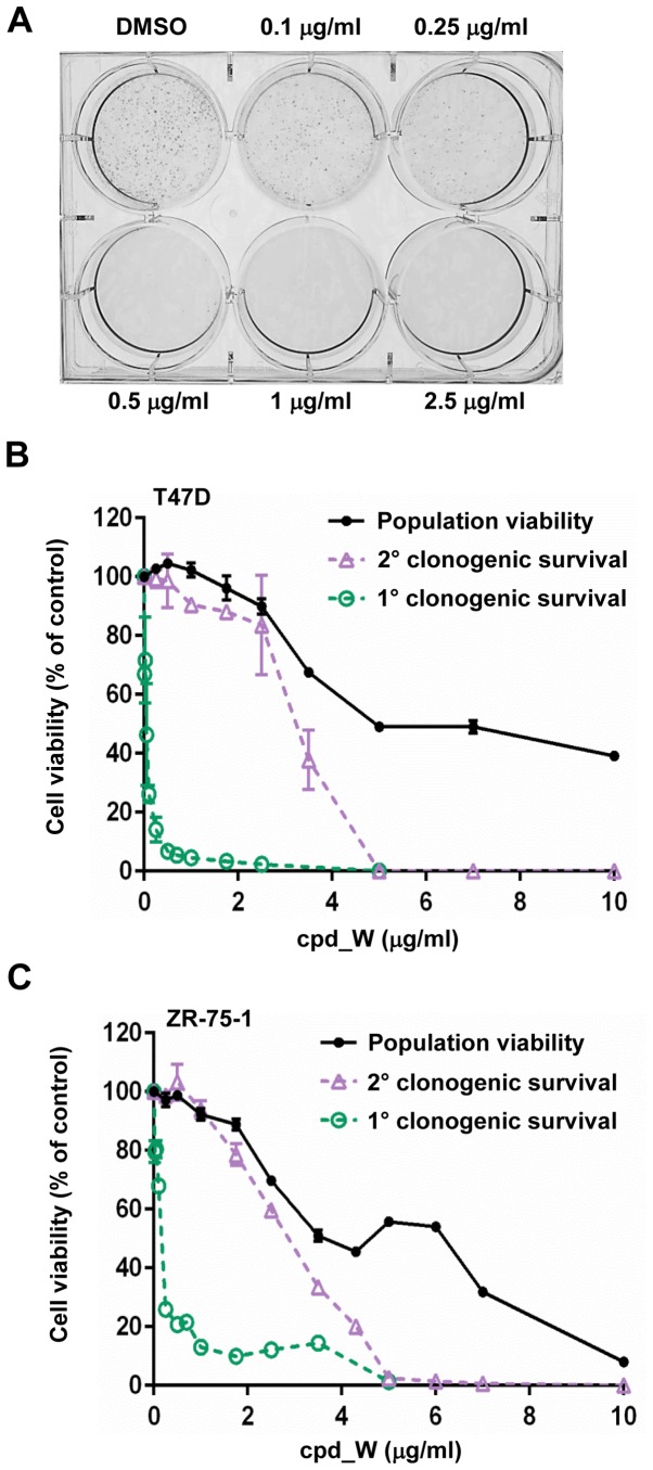
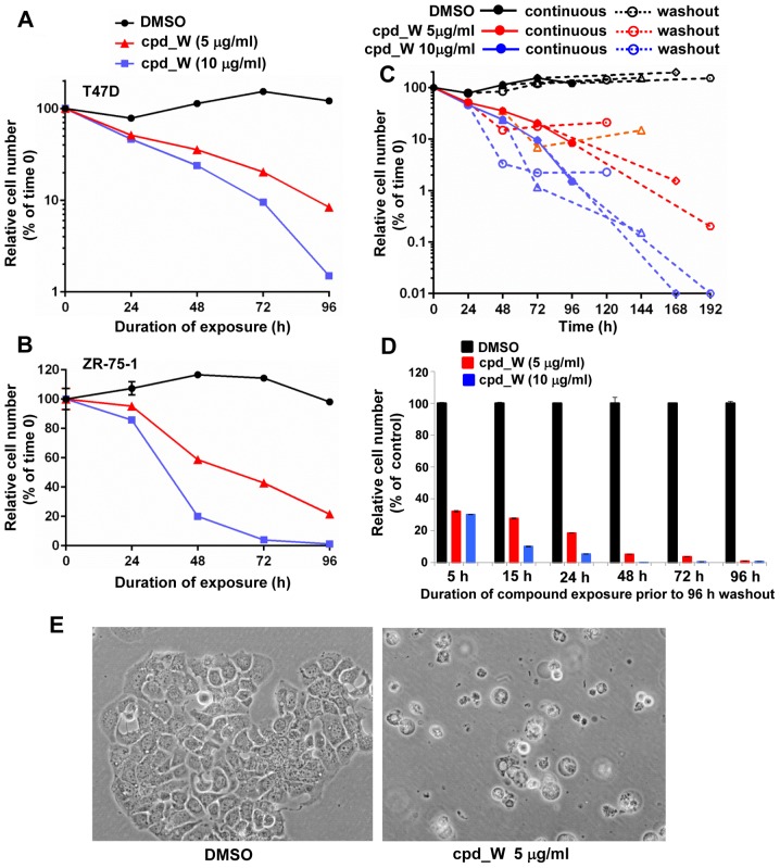
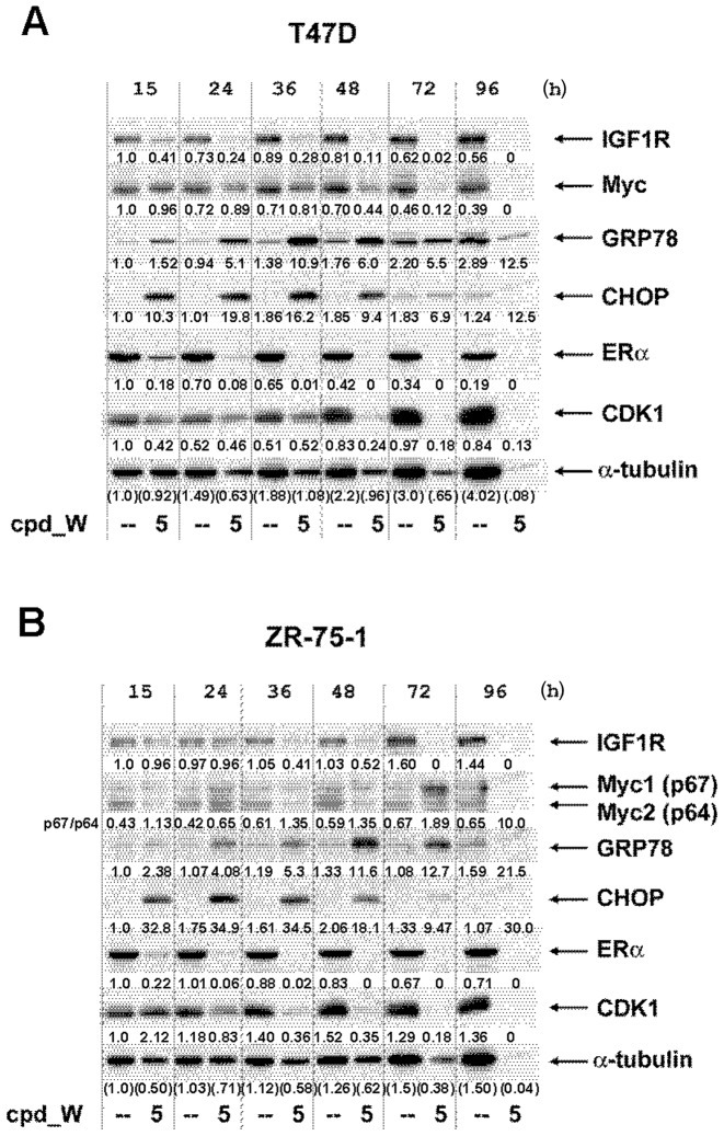
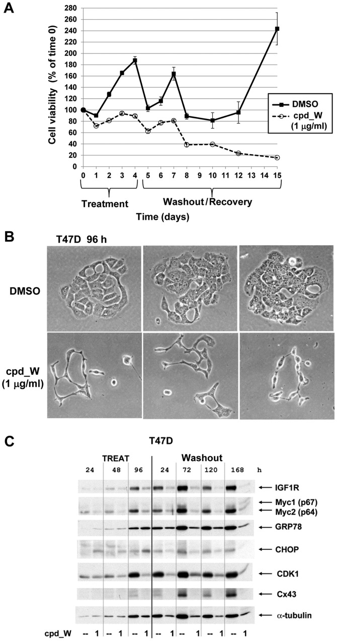
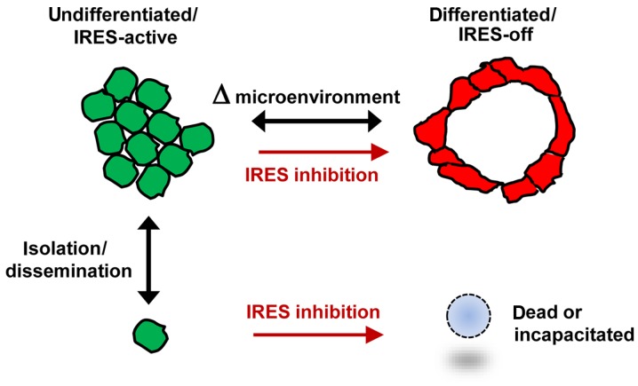
Using a series of potential biomarkers relevant to mechanisms of protein synthesis, we observed that estrogen receptor (ER)-positive breast tumor cells exist in two distinct yet interconvertible phenotypic states (of roughly equal proportion) which differ in the degree of differentiation and use of IRES-mediated translation. Nascently translated IGF1R in the cytoplasm positively correlated with IRES activity and the undifferentiated phenotype, while epitope accessibility of RACK1, an integral component of the 40S ribosomal subunit, aligned with the more differentiated IRES-off state. When deprived of soluble growth factors, the entire tumor cell population shifted to the undifferentiated phenotype in which IRES-mediated translation was active, facilitating survival under these adverse microenvironmental conditions. However, if IRES-mediated translation was inhibited, the cells instead were forced to transition uniformly to the more differentiated state. Notably, cytoplasmic localization of estrogen receptor α (ERα/ESR1) precisely mirrored the pattern observed with nascent IGF1R, correlating with the undifferentiated IRES-active phenotype. Inhibition of IRES-mediated translation resulted in both a shift in ERα to the nucleus (consistent with differentiation) and a marked decrease in ERα abundance (consistent with the inhibition of ERα synthesis via its IRES). Although breast tumor cells tolerated forced differentiation without extensive loss of their viability, their reproductive capacity was severely compromised. In addition, CDK1 was decreased, connexin 43 eliminated and Myc translation altered as a consequence of IRES inhibition. Isolated or low-density ER-positive breast tumor cells were particularly vulnerable to IRES inhibition, losing the ability to generate viable cohesive colonies, or undergoing massive cell death. Collectively, these results provide further evidence for the integral relationship between IRES-mediated translation and the undifferentiated phenotype and demonstrate how therapeutic manipulation of this specialized mode of protein synthesis may be used to limit the phenotypic plasticity and incapacitate or eliminate these otherwise highly resilient breast tumor cells.


Figures

Figure 1.
Figure 1.
IRES-mediated translation and IRES inhibition in a cell-free system and in cells. (A) Diagrammatic comparison of general protein synthesis to IRES-mediated translation. General protein synthesis is mediated by cap-dependent ribosomal scanning from the 5′-end of the mRNA and may be modulated by mTOR inhibitors. Internal ribosome entry sites (IRESs) allow the 40S ribosome to engage the mRNA at a position much closer (in many cases immediately adjacent to) the AUG initiation codon. IRES-mediated translation is independently regulated and serves as a fail-safe mechanism ensuring the synthesis of proteins most critical for cell survival. (B) Structure of IRES inhibitor lead compound W (cpd_W): Ethyl 2-{[2-(1,3-benzoxazol-2-ylthio)butanoyl]amino}-4-methyl-1,3-thiazole-5-carboxylate, MW 405. (C) In vitro translation assays: Rabbit reticulocyte lysate was programmed with a bicistronic reporter RNA in which translation of the second cistron (firefly luciferase coding sequence) is mediated by the IGF1R IRES, while translation of the first cistron (Renilla luciferase coding sequence) is mediated by ribosomal scanning. IRES inhibitor cpd_W (or vehicle control) was included in the reaction in increasing concentrations as indicated. The result is indicative of selective inhibition of IRES-mediated translation. A structural analog of cpd_W (W-7) in which a single atom has been modified (converting the benzoxazole to a benzimidazole) was completely inactive in this assay, indicative of the chemical specificity of IRES inhibition. Cycloheximide (5 µg/ml, chx) and puromycin (250 µg/ml, puro) were included as reference standards for non-specific translational inhibition (far right). (D) IRES inhibitor cpd_W completely blocked de novo synthesis of IGF1R in breast tumor cells under adverse conditions (serum-deprivation, loss of adhesion) relevant to the microenvironment of the tumor. T47D breast tumor cells were seeded in 6-well plates and allowed 48 h to recover and resume proliferation, then incubated in the presence of IRES inhibitor cpd_W (10 µg/ml) or vehicle control (0.1% DMSO) as indicated. The cells were simultaneously subjected to acute serum deprivation (0.5% fetal calf serum, no added insulin) to increase dependence on IRES-mediated translation. After 24 h, the cells were harvested and whole cell lysates prepared, equivalent aliquots separated by SDS-PAGE and immunoblotted for IGF1R-β and α-tubulin. In lanes 7–12, the cells were trypsinized and seeded into 6-well plates and immediately incubated in the presence of IRES-inhibitor cpd_W or vehicle control as indicated. Robust regeneration of trypsin-catabolized IGF1R was observed within 24 h in vehicle-treated cells, however, this was completely blocked in the presence of cpd_W (10 µg/ml as shown; IC50, 2 µg/ml). The asterisk (*) marks the position of trypsin-catabolized IGF1R. In lanes 13–17, the cells were treated as described for lanes 7–12, except that following trypsinization, cells were transferred to low-adherence plates, forcing cells to adapt to a state of anchorage-independence. The results confirmed the activity of cpd_W against the endogenous IRES in genetically-unmodified tumor cells. Similar results were obtained with IRES inhibitor lead cpd_P (11).
Figure 2.
Figure 2.
ER-positive breast tumor cell transition between two distinct phenotypic states distinguished by differentiation status and IRES-mediated translation. T47D breast tumor cells were seeded in 8-well chamber slides, allowed 48 h to recover and resume proliferation, then subjected to acute serum deprivation (0.5% FCS) in the presence of cpd_W at 10 µg/ml or vehicle control (0.1% DMSO). At the indicated time-points, the cells were fixed, permeabilized and stained using N20 antibody to IGF1R and clone 20 antibody to RACK1, as described in the Materials and methods section. (A) Confocal images illustrating two distinct populations of tumor cells distinguished by mutually exclusive IGF1R N20 and RACK1 staining patterns. (B) RACK1/IGF1R N20 images of cells at 10, 34 or 58 h time-points, demonstrated progressive phenotypic transitions. Cells subjected to serum deprivation (top row) became almost universally IGF1R N20-positive, while cells treated with IRES inhibitor cpd_W under the same conditions (bottom row) became entirely RACK1-positive. (C) Higher magnification image from cpd_W 10 h field in which cells bearing distinct cytoplasmic RACK1-positive foci (circled) were demonstrated. (D) Higher magnification image from DMSO 10 h field in which biphenotypic cells (circled) were demonstrated.
Figure 3.
Figure 3.
Biomarkers of the undifferentiated/IRES-active and differentiated/IRES-off phenotypes. (A) T47D cells were treated with IRES inhibitor cpd_W as described in Fig. 2. At the 48 h time-point, the cells were fixed and stained for ERα (D8H8) and RACK1. (B) T47D cells were treated with cpd_W for 15, 24 or 48 h. Exposure to cpd_W was terminated by replacing with fresh media and cells were fixed and stained for ERα and RACK1 at the 48 h time-point. (C) ZR-75-1 cells were treated and stained for ERα and RACK1 as abovedescribed in (A). (D-J) T47D (D, F, H and J) or ZR-75-1 (E, G and I) cells were treated with cpd_W or vehicle control for 48 h, then stained for IGF1R N20 and GRP78 (D and E), RACK1 and ZO-1 (F and G), RACK1 and CLIMP-63 (H), ERα and E-cadherin (I), or IGF1R N20 and α-tubulin (J).
Figure 3.
Figure 3.
Biomarkers of the undifferentiated/IRES-active and differentiated/IRES-off phenotypes. (A) T47D cells were treated with IRES inhibitor cpd_W as described in Fig. 2. At the 48 h time-point, the cells were fixed and stained for ERα (D8H8) and RACK1. (B) T47D cells were treated with cpd_W for 15, 24 or 48 h. Exposure to cpd_W was terminated by replacing with fresh media and cells were fixed and stained for ERα and RACK1 at the 48 h time-point. (C) ZR-75-1 cells were treated and stained for ERα and RACK1 as abovedescribed in (A). (D-J) T47D (D, F, H and J) or ZR-75-1 (E, G and I) cells were treated with cpd_W or vehicle control for 48 h, then stained for IGF1R N20 and GRP78 (D and E), RACK1 and ZO-1 (F and G), RACK1 and CLIMP-63 (H), ERα and E-cadherin (I), or IGF1R N20 and α-tubulin (J).
Figure 4.
Figure 4.
Loss of reproductive capacity in ER-positive breast tumor cells as a consequence of inhibition of IRES-mediated translation. T47D (A) or ZR-75-1 (B) cells ~75% confluent were treated with IRES inhibitor cpd_W at concentrations ranging from 0.25–10 µg/ml for 96 h, after which the viability was assessed based on ATP content as described in Materials and methods section. The results (population viability, black lines) are plotted relative to vehicle control. In parallel, a set of identically prepared samples, following 96 h treatment, was provided fresh media (containing full serum) and allowed to recover for 24 h, then trypsinized and reseeded at low density, allowed 6 days to proliferate, then viability assessed as a measure of reproductive capacity (secondary clonogenic survival, purple lines).
Figure 5.
Figure 5.
Impact of IRES inhibition on IGF1R, Myc, ERα, CDK1 and connexin 43 (Cx43). T47D (A) or ZR-75-1 (B) cells ~75% confluent were treated with varying concentrations of IRES inhibitor cpd_W or vehicle control as indicated for 24 or 48 h, or 48 h followed by a 24 h washout period in media with no compound added. Whole cell lysates were prepared, equivalent aliquots separated by SDS-PAGE and used for western blot analyses of the indicated proteins.
Figure 6.
Figure 6.
Loss of clonogenic survival in ER-positive breast tumor cells subjected to inhibition of IRES-mediated translation. (A) T47D cells were seeded at low (clonogenic) density (~700 cells/cm2), allowed 48 h to recover and then treated with IRES inhibitor cpd_W at increasing concentrations as indicated for 96 h under low serum (0.5% FCS) conditions, followed by 11 days of incubation in absence of compound. Colonies were stained with MTT to enhance visualization. T47D (B) or ZR-75-1 (C) cells were seeded at clonogenic density in 24 well plates, allowed 48 h to recover, then treated with cpd_W (or vehicle control, DMSO 0.1%) under low serum conditions for 96 h, after which fresh media was provided and the cells were allowed 9–10 days to proliferate. Cell viability was assessed and used as a quantitative surrogate for colony formation. The results (primary clonogenic survival, green lines) are superimposed on the graphs from Fig. 4, to facilitate direct comparisons. The use of ATP-based assay tended to underestimate the negative impact of IRES inhibition on clonogenic survival, since cells may register as viable yet not be capable of generating a colony.
Figure 7.
Figure 7.
Massive cell death as a consequence of IRES inhibition in low-density ER-positive breast tumor cells. T47D (A) or ZR-75-1 (B) cells were plated at clonogenic density, allowed 48 h to recover, then treated with IRES inhibitor cpd_W at 5 or 10 µg/ml or vehicle control under low serum conditions for up to 96 h and then viability was assessed at 24 h intervals. (C) T47D cells were seeded at clonogenic density then treated with cpd_W at 5 µg/ml (red lines) or 10 µg/ml (blue lines) or vehicle (DMSO) control (black lines) for 24, 48, 72 or 96 h. The cells were then provided fresh media without compound and allowed 96 h to recover and proliferate before viability was again assessed. Solid symbols and lines are indicative of continuous treatment, while open symbols and dashed lines are indicative of washout period. The trajectories of the dashed lines allowed us to gauge the impact of IRES inhibition on cell survival following removal of the compound. (D) ZR-75-1 cells were seeded at clonogenic density then treated with cpd_W at 5 or 10 µg/ml for 5, 15, 24, 48, 72 or 96 h, after which all samples were provided fresh media and allowed 96 h to recover in the absence of compound. The graph plotted relative viability at the end of the 96 h washout period. (E) Phase contrast images of T47D cells treated at low density with cpd_W at 5 µg/ml for 96 h.
Figure 8.
Figure 8.
Enhanced sensitivity of IGF1R, Myc, ERα and CDK1 to IRES inhibition in low-density ER-positive breast tumor cells. T47D (A) or ZR-75-1 (B) cells were seeded at clonogenic density in groups of three replicate 10 cm2 wells and treated with cpd_W at 5 µg/ml or vehicle control under low serum conditions for up to 96 h. Whole cell lysates were combined from three wells of each condition at each time-point. Equivalent aliquots (by volume, not protein content) were electrophoresed on 10 or 13% polyacrylamide gels and used for western blot analysis of the indicated proteins. Variations in intensity of α-tubulin bands are indicative of changes in viable cell number during the course of the experiment. The light grey vertical lines placed over the ECL images were included only for orientation; the 12 lanes are contiguous. The numbers superimposed below each band are indicative of relative intensity as determined by quantitative densitometry. Values for α-tubulin (in parentheses) were expressed relative to the vehicle control at the earliest time-point. Values for other proteins were normalized to the abundance of α-tubulin in each sample. Values for Myc in ZR-75-1 cells represented the ratio of intensities of the p67 and p64 isoforms, which were reversed as a consequence of treatment with the IRES inhibitor.
Figure 9.
Figure 9.
ER-positive breast tumor cells lose the ability to generate viable cohesive colonies when exposed to very low concentrations of IRES inhibitor cpd_W. (A) T47D cells were seeded at clonogenic density, treated for 96 h with cpd_W at 1 µg/ml or vehicle control (0.01% DMSO), then provided fresh media and allowed 11 days for recovery. Low serum conditions were maintained throughout the experiment. The viability was assessed periodically on replicate wells throughout the course of the experiment. (B) Phase contrast images captured following 96 h treatment of low-density T47D cells with 1 µg/ml cpd_W revealed the defect in colony formation induced by very low concentration of the IRES inhibitor. (C) Western blot analysis of T47D cells treated at clonogenic density with IRES inhibitor cpd_W at 1 µg/ml as abovedescribed in (A). Whole cell lysates were collected as described in the legend of Fig. 8. Cx43, connexin 43.
Figure 10.
Figure 10.
Relationship between differentiation status and IRES-mediated translation in ER-positive breast tumor cells. A working model based on the collective results of the experiments in the present study. ER-positive breast tumor cells transition between two distinct phenotypes: i) an undifferentiated state (green), which is absolutely dependent upon IRES-mediated translation and associated with extraordinary resiliency toward adverse microenvironmental conditions; and ii) a moderately differentiated state (red), in which IRES-mediated translation is not active and tumor cells display features of normal epithelial cells. The transition between these two phenotypes is reversible and this plasticity contributes to therapeutic failure and progressive disease. However, chemical IRES inhibition forces tumor cells to differentiate and this forced transition may become irreversible (terminal), with loss of reproductive capacity. Isolated/low-density ER-positive breast tumor cells depend on IRES-mediated translation and the undifferentiated phenotype to survive with limited paracrine support and no intercellular contact. These otherwise highly resilient tumor cells are exquisitely sensitive to IRES inhibition.

Similar articles

Cited by

References

    1. Prats AC, Prats H. Translational control of gene expression: Role of IRESs and consequences for cell transformation and angiogenesis. Prog Nucleic Acid Res Mol Biol. 2002;72:367–413. doi: 10.1016/S0079-6603(02)72075-8. - DOI - PubMed
    1. Baird SD, Turcotte M, Korneluk RG, Holcik M. Searching for IRES. RNA. 2006;12:1755–1785. doi: 10.1261/rna.157806. - DOI - PMC - PubMed
    1. Spriggs KA, Stoneley M, Bushell M, Willis AE. Re-programming of translation following cell stress allows IRES-mediated translation to predominate. Biol Cell. 2008;100:27–38. doi: 10.1042/BC20070098. - DOI - PubMed
    1. Meng Z, Jackson NL, Choi H, King PH, Emanuel PD, Blume SW. Alterations in RNA-binding activities of IRES-regulatory proteins as a mechanism for physiological variability and pathological dysregulation of IGF-IR translational control in human breast tumor cells. J Cell Physiol. 2008;217:172–183. doi: 10.1002/jcp.21486. - DOI - PubMed
    1. Nagamachi A, Htun PW, Ma F, Miyazaki K, Yamasaki N, Kanno M, Inaba T, Honda Z, Okuda T, Oda H, et al. A 5′untranslated region containing the IRES element in the Runx1 gene is required for angiogenesis, hematopoiesis and leukemogenesis in a knock-in mouse model. Dev Biol. 2010;345:226–236. doi: 10.1016/j.ydbio.2010.07.015. - DOI - PubMed