. 2018 Jul;42(1):346-358.
doi: 10.3892/ijmm.2018.3606. Epub 2018 Mar 30.

TIM‑4 blockade of KCs combined with exogenous TGF‑β injection helps to reverse acute rejection and prolong the survival rate of mice receiving liver allografts

Affiliations

TIM‑4 blockade of KCs combined with exogenous TGF‑β injection helps to reverse acute rejection and prolong the survival rate of mice receiving liver allografts

Hao Wu et al. Int J Mol Med. 2018 Jul.

Abstract

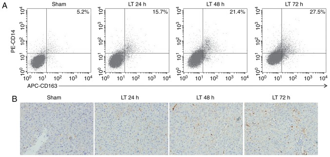
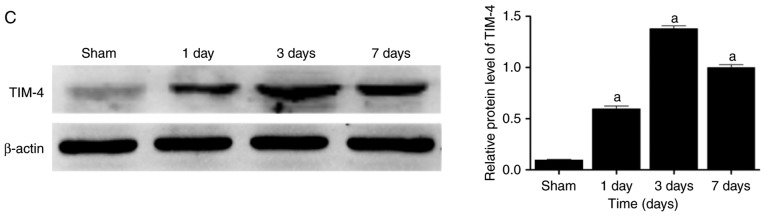
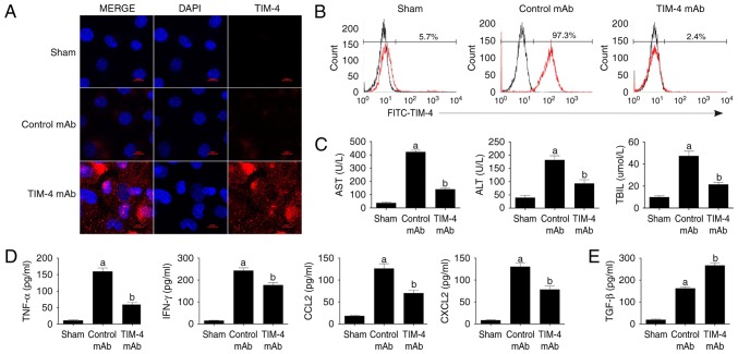
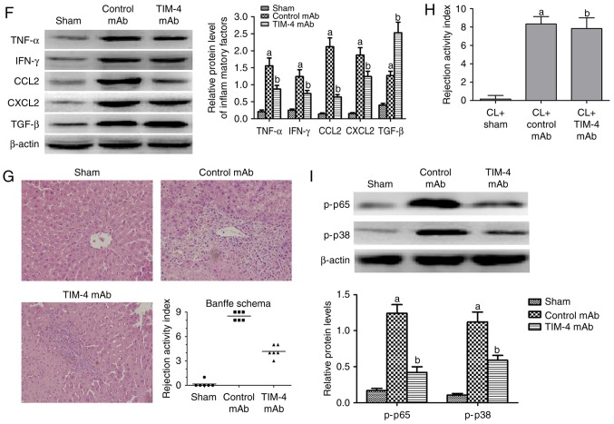
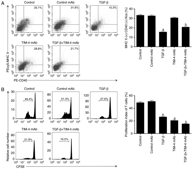
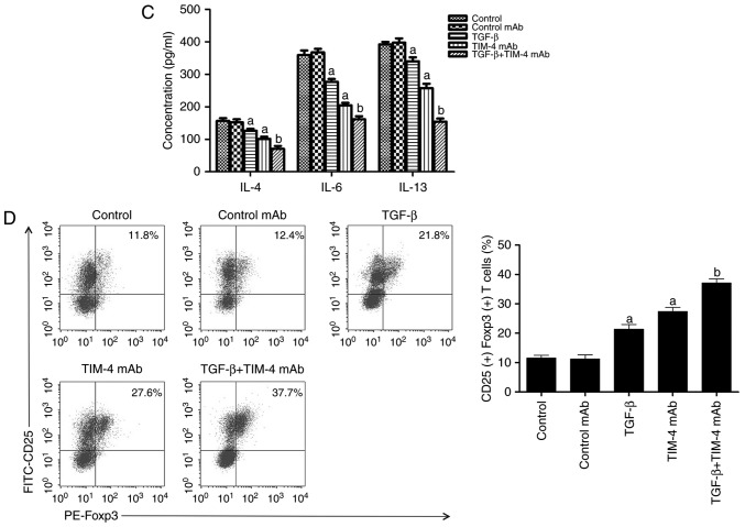
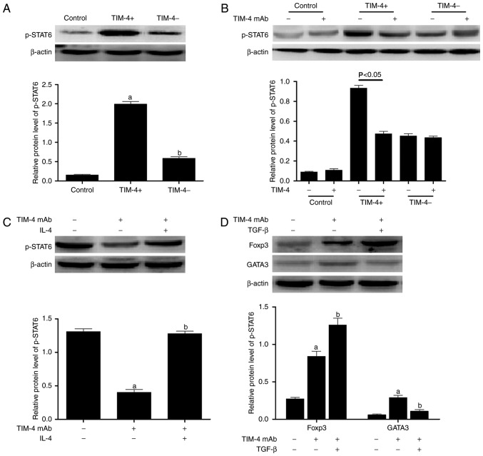
An acute reaction response (AR) following liver transplantation (LT) is caused by immune responses that are primarily mediated by T lymphocytes. Kupffer cells (KCs) are the largest antigen presenting cell (APC) group in vivo and are the primary modulators of the inflammatory or tolerogenic immune response in liver tissues. T cell immunoglobulin‑domain and mucin‑domain-4 (TIM‑4), the only TIM protein not expressed on T cells, is expressed on APCs; suggesting that it mediates the various immune responses. However, to the best of our knowledge, the role of TIM‑4 expressed by KCs in LT injury remains unknown. The present study aimed to explore whether and how TIM‑4 expressed by KCs is involved in the AR of liver allografts. Orthotopic liver transplantation (OLT) was performed in mice to establish a model of AR and results demonstrated that LT may lead to the augmented expression of TIM‑4 in activated KCs. It was also revealed that TIM‑4 blockade markedly attenuated AR injury in vivo via the nuclear factor‑κB (NF‑κB) and p38 mitogen‑activated protein kinase (p38 MAPK) signaling pathways. In addition, levels of transforming growth factor‑β (TGF‑β) were increased following TIM‑4 blockade. Furthermore, in a KC/cluster of differentiation (CD)4+ T cell co‑culture system, blocking TIM‑4 inhibited T helper 2 (Th2) differentiation, stimulated the conversion of naive (CD)4+ T cells into CD4+CD25+Forkhead box protein p3+ T regulatory cells and suppressed interleukin‑4/signal transducer and activator of transcription 6/transcription factor gata3 signaling. These effects were enhanced following the addition of TGF‑β. It was also demonstrated that LT mouse models treated with TIM‑4 blockade in combination with exogenous TGF‑β injections, increased the survival times of mice and enhanced the amelioration of AR in LT. These results indicate that blocking the expression of TIM‑4 by KCs via exogenous TGF‑β injection may be an effective therapeutic strategy to inhibit the AR of liver allografts.


Figures

Figure 1
Figure 1
Orthotopic liver transplantation increases KC activation and TIM-4 expression. (A) KCs were isolated from mice in the sham and LT groups 24, 48 and 72 h following surgery. Cells were double stained with PE-CD14 and APC-CD163 and quantified using flow cytometry. (B) Liver samples were obtained from mice in the sham and LT groups and assessed using immunohistochemistry (magnification, ×400). Activated hepatic KCs are stained brown. (C) KCs were isolated from mice in the sham and LT groups 1, 3 and 7 days following surgery and TIM-4 expression was analyzed using western blotting. Data are presented as the mean ± standard deviation. aP<0.05 vs. sham group. KC, kupffer cells; TIM-4, T cell immunoglobulin-domain mucin-domain-4; CD, cluster of differentiation; APC, antigen presenting cell; LT, liver transplantation; PE, phycoerythrin; d, days.
Figure 1
Figure 1
Orthotopic liver transplantation increases KC activation and TIM-4 expression. (A) KCs were isolated from mice in the sham and LT groups 24, 48 and 72 h following surgery. Cells were double stained with PE-CD14 and APC-CD163 and quantified using flow cytometry. (B) Liver samples were obtained from mice in the sham and LT groups and assessed using immunohistochemistry (magnification, ×400). Activated hepatic KCs are stained brown. (C) KCs were isolated from mice in the sham and LT groups 1, 3 and 7 days following surgery and TIM-4 expression was analyzed using western blotting. Data are presented as the mean ± standard deviation. aP<0.05 vs. sham group. KC, kupffer cells; TIM-4, T cell immunoglobulin-domain mucin-domain-4; CD, cluster of differentiation; APC, antigen presenting cell; LT, liver transplantation; PE, phycoerythrin; d, days.
Figure 2
Figure 2
Blockade of TIM-4 improves hepatic acute reaction response injury and reduces the secretion of inflammatory cytokines. (A) KCs were isolated from mice in the sham, control mAb and TIM-4 mAb groups on day 7 post-surgery. Cells were stained with specific secondary antibodies labeled with tetramethylrhodamine (red) and DAPI (blue) and then examined using laser confocal microscopy (magnification ×800). (B) KCs were isolated from each group and stained with FITC-TIM-4 mAb for flow cytometry. (C) Blood samples were obtained (0.5 ml/mouse) from murine abdominal aortas in each group on day 7 post-surgery. Levels of AST, ALT and TBIL were determined in a clinical biochemical laboratory. (D-F) Levels of inflammatory cytokines, including TNF-α, IFN-γ, CCL2, CXCL2 and TGF-β were detected using ELISA and western blotting. (G) Representative micrographs of the pathological damage observed in each group post-transplantation following staining with hematoxylin and eosin (magnification ×400). (H) Donor mice received CL treatment to destroy KCs prior to surgery. orthotopic liver transplantation or sham surgery was subsequently performed and mice were treated either with or without TIM-4 mAb. All mice underwent analysis to determine the RAI score on day 7 following surgery. (I) The expression of key phosphoproteins involved in the NF-κB and p38 MAPK signaling pathways, including p65 and p38, respectively, were analyzed using western blotting. Data are presented as the mean ± standard deviation. aP<0.05 vs. sham group; bP<0.05 vs. control mAb group. TIM-4, T cell immunoglobulin-domain mucin-domain-4; mAb, monoclonal antibodies; KCs, kupffer cells; FITC, fluorescein isothiocyanate; AST, aspartate aminotransferase; ALT, alanine aminotransferase; TBIL, total bilirubin in serum; TNF-α, tumor necrosis factor-α; IFN-γ, interferon-γ; CCL2, chemokine ligand ; CXCL2, C-X-C motif chemokine ligand 2; TGF-β, transforming growth factor-β; CL, clondronate liposomes; NF-κB, nuclear factor-κB; MAPK, mitogen-activated protein kinase.
Figure 2
Figure 2
Blockade of TIM-4 improves hepatic acute reaction response injury and reduces the secretion of inflammatory cytokines. (A) KCs were isolated from mice in the sham, control mAb and TIM-4 mAb groups on day 7 post-surgery. Cells were stained with specific secondary antibodies labeled with tetramethylrhodamine (red) and DAPI (blue) and then examined using laser confocal microscopy (magnification ×800). (B) KCs were isolated from each group and stained with FITC-TIM-4 mAb for flow cytometry. (C) Blood samples were obtained (0.5 ml/mouse) from murine abdominal aortas in each group on day 7 post-surgery. Levels of AST, ALT and TBIL were determined in a clinical biochemical laboratory. (D-F) Levels of inflammatory cytokines, including TNF-α, IFN-γ, CCL2, CXCL2 and TGF-β were detected using ELISA and western blotting. (G) Representative micrographs of the pathological damage observed in each group post-transplantation following staining with hematoxylin and eosin (magnification ×400). (H) Donor mice received CL treatment to destroy KCs prior to surgery. orthotopic liver transplantation or sham surgery was subsequently performed and mice were treated either with or without TIM-4 mAb. All mice underwent analysis to determine the RAI score on day 7 following surgery. (I) The expression of key phosphoproteins involved in the NF-κB and p38 MAPK signaling pathways, including p65 and p38, respectively, were analyzed using western blotting. Data are presented as the mean ± standard deviation. aP<0.05 vs. sham group; bP<0.05 vs. control mAb group. TIM-4, T cell immunoglobulin-domain mucin-domain-4; mAb, monoclonal antibodies; KCs, kupffer cells; FITC, fluorescein isothiocyanate; AST, aspartate aminotransferase; ALT, alanine aminotransferase; TBIL, total bilirubin in serum; TNF-α, tumor necrosis factor-α; IFN-γ, interferon-γ; CCL2, chemokine ligand ; CXCL2, C-X-C motif chemokine ligand 2; TGF-β, transforming growth factor-β; CL, clondronate liposomes; NF-κB, nuclear factor-κB; MAPK, mitogen-activated protein kinase.
Figure 3
Figure 3
The effect of TIM-4 blockade with addition of exogenous TGF-β on inhibiting T helper 2 cell differentiation and inducing the conversion of CD4+CD25+Foxp3+ T regulatory cells. (A) TIM-4+ KCs were isolated from model mice and purified using fluorescence activated cell sorting, followed by addition of control mAb/TGF-β/TIM-4 mAb/TGF-β+TIM-4 mAb into medium for incubation. Following washing and resuspension of cells, they were stained with PEcy5-MHC II and PE-CD40 antibodies and acquired for FACS analysis. (B) Spleen CD4+CD25 T cells were purified from recipient mice and labeled with CFSE, followed by co-culturing with KCs in condition described in A (control as untreated). The proliferation of T cells was then determined using a CFSE dilution gated on CD4+ populations. (C) The supernatants of the co-cultured system, including the cytokines IL-4, IL-6 and IL-13, were detected using enzyme-linked immunosorbent assay. (D) Splenic CD4+CD25 T cells were purified from recipient mice and co-cultured with KCs in conditions as described in A. Following washing and re-suspension, the cells were stained with PE-Foxp3 and FITC-CD25 antibodies and underwent FACS analysis. Data are presented as the mean ± standard deviation. aP<0.05 vs. control and control mAb groups; bP<0.05 vs. TGF-β and TIM-4 mAb groups. TIM-4, T cell immunoglobulin-domain mucin-domain-4; TGF-β, transforming growth factor-β; KCs, kupffer cells; mAb, monoclonal antibodies; MHC II, major histocompatibility complex II; CD, cluster of differentiation; FACS, fluorescence-activated cell sorting; CFSE, carboxyfluorescein succinimidyl ester; IL, interleukin; Foxp3, forkhead box P3; FITC, fluorescein isothiocyanate; PE, phycoerythrin; PEcy5, phycoerythrin-streptavidin.
Figure 3
Figure 3
The effect of TIM-4 blockade with addition of exogenous TGF-β on inhibiting T helper 2 cell differentiation and inducing the conversion of CD4+CD25+Foxp3+ T regulatory cells. (A) TIM-4+ KCs were isolated from model mice and purified using fluorescence activated cell sorting, followed by addition of control mAb/TGF-β/TIM-4 mAb/TGF-β+TIM-4 mAb into medium for incubation. Following washing and resuspension of cells, they were stained with PEcy5-MHC II and PE-CD40 antibodies and acquired for FACS analysis. (B) Spleen CD4+CD25 T cells were purified from recipient mice and labeled with CFSE, followed by co-culturing with KCs in condition described in A (control as untreated). The proliferation of T cells was then determined using a CFSE dilution gated on CD4+ populations. (C) The supernatants of the co-cultured system, including the cytokines IL-4, IL-6 and IL-13, were detected using enzyme-linked immunosorbent assay. (D) Splenic CD4+CD25 T cells were purified from recipient mice and co-cultured with KCs in conditions as described in A. Following washing and re-suspension, the cells were stained with PE-Foxp3 and FITC-CD25 antibodies and underwent FACS analysis. Data are presented as the mean ± standard deviation. aP<0.05 vs. control and control mAb groups; bP<0.05 vs. TGF-β and TIM-4 mAb groups. TIM-4, T cell immunoglobulin-domain mucin-domain-4; TGF-β, transforming growth factor-β; KCs, kupffer cells; mAb, monoclonal antibodies; MHC II, major histocompatibility complex II; CD, cluster of differentiation; FACS, fluorescence-activated cell sorting; CFSE, carboxyfluorescein succinimidyl ester; IL, interleukin; Foxp3, forkhead box P3; FITC, fluorescein isothiocyanate; PE, phycoerythrin; PEcy5, phycoerythrin-streptavidin.
Figure 4
Figure 4
TIM-4 blockade stimulates the differentiation of T cells into CD4+CD25+Foxp3+ T regulatory cells via the IL-4/STAT6/Gata3 pathway. (A) TIM-4+ and TIM-4 KCs were obtained from mice following liver transplantation and co-cultured with naive CD4+ T cells. The expression of p-STAT6 in T cells was determined using western blotting. (B) TIM-4 mAb was added to co-cultured cohorts as described in A, to analyze the expression of p-STAT6 in T cells. (C) The exogenous addition of IL-4 to TIM-4+ KCs co-cultured with naive CD4+ T cells (that received either no pretreatment or pretreatment with TIM-4 mAb) was performed and the expression of p-STAT6 in T cells was analyzed. (D) TGF-β was added to TIM-4+ KCs co-cultured with naive CD4+ T cells (that received either no pretreatment or pretreatment with TIM-4 mAb) to determine the expression of Foxp3 and Gata3 protein in T cells. Data are presented as the mean ± standard deviation of the mean. aP<0.05 vs. control; bP<0.05 vs. TIM-4+ group. TIM-4, T cell immunoglobulin-domain mucin-domain-4; IL-4, interleukin-4; p-, phosphorylated; STAT6, signal transducer and activator of transcription 6; Gata3, transcription factor gata3; CD, cluster of differentiation; mAb, monoclonal antibodies; KCs, kupffer cells; TGF-β, transforming growth factor-β; Foxp3, forkhead box P3.
Figure 5
Figure 5
The effect of TIM-4 blockade with exogenous TGF-β injection on acute reaction response in vivo. (A) Mice were injected with either anti-TIM-4 mAb (0.35 mg/mouse), 0.5 ml TGF-β (1 ng/ml) or in combination via murine portal veins to establish LT (LT group were treated with PBS as control). Treatment in each group was administered continuously at the same dose for a total of 2 days following surgery. Levels of AST, ALT and TBIL were determined in a clinical biochemical laboratory on day 7 following surgery. (B) Hepatocyte apoptosis was detected using the terminal deoxynucleotidyl transferase-mediated 2′-deoxyuridine 5′-triphosphate nick-end labeling method (magnification ×400). (C) T cells were purified from hepatic tissue. Following washing and re-suspension, cells were stained with PE-Foxp3 and FITC-CD25 antibodies and acquired for fluorescence-activated cell sorting analysis. (D) Mice survival time was observed and analyzed using the Log-rank test. Data are presented as the mean ± standard deviation of the mean. aP<0.05 vs. LT group; bP<0.05 vs. TGF-β and TIM-4 mAb groups. TIM-4, T cell immunoglobulin-domain mucin-domain-4; TGF-β, transforming growth factor-β; mAb, monoclonal antibodies; LT, liver transplantation; AST, aspartate aminotransferase; ALT, alanine aminotransferase; TBIL, total bilirubin in serum; Foxp3, forkhead box P3; FITC, fluorescein isothiocyanate; CD, cluster of differentiation; PE, phycoerythrin.

References

    1. Knechtle SJ, Kwun J. Unique aspects of rejection and tolerance in liver transplantation. Semin Liver Dis. 2009;29:91–101. doi: 10.1055/s-0029-1192058. - DOI - PubMed
    1. Yoshida O, Dou L, Kimura S, Yokota S, Isse K, Robson SC, Geller DA, Thomson AW. CD39 deficiency in murine liver allografts promotes inflammatory injury and immune-mediated rejection. Transpl Immunol. 2015;32:76–83. doi: 10.1016/j.trim.2015.01.003. - DOI - PMC - PubMed
    1. Shen ZY, Wu B, Liu T, Yang Y, Yin ML, Zheng WP, Zhang BY, Song HL. Immunomodulatory effects of bone marrow mesenchymal stem cells overexpressing heme oxygenase-1: Protective effects on acute rejection following reduced-size liver transplantation in a rat model. Cell Immunol. 2017;313:10–24. doi: 10.1016/j.cellimm.2016.12.006. - DOI - PubMed
    1. Meyers JH, Sabatos CA, Chakravarti S, Kuchroo VK. The TIM gene family regulates innate and adaptive immunity. Trends Mol Med. 2005;11:362–269. doi: 10.1016/j.molmed.2005.06.008. - DOI - PubMed
    1. Freeman GJ, Casasnovas JM, Umetsu DT, DeKruyff RH. TIM genes: A family of cell surface phosphatidylserine receptors that regulate innate and adaptive immunity. Immunol Rev. 2010;235:172–189. doi: 10.1111/j.0105-2896.2010.00903.x. - DOI - PMC - PubMed

MeSH terms