. 2018 Jun;39(6):2537-2544.
doi: 10.3892/or.2018.6342. Epub 2018 Mar 30.

Enrichment and mutation detection of circulating tumor cells from blood samples

Affiliations

Enrichment and mutation detection of circulating tumor cells from blood samples

Ruqin Kou et al. Oncol Rep. 2018 Jun.

Abstract

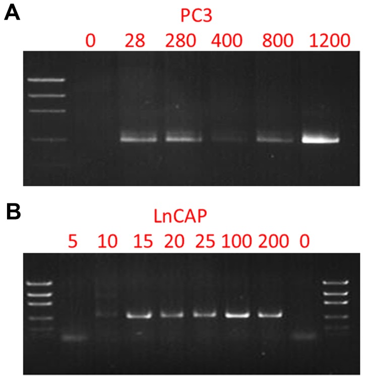
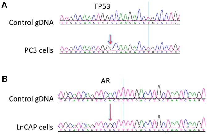
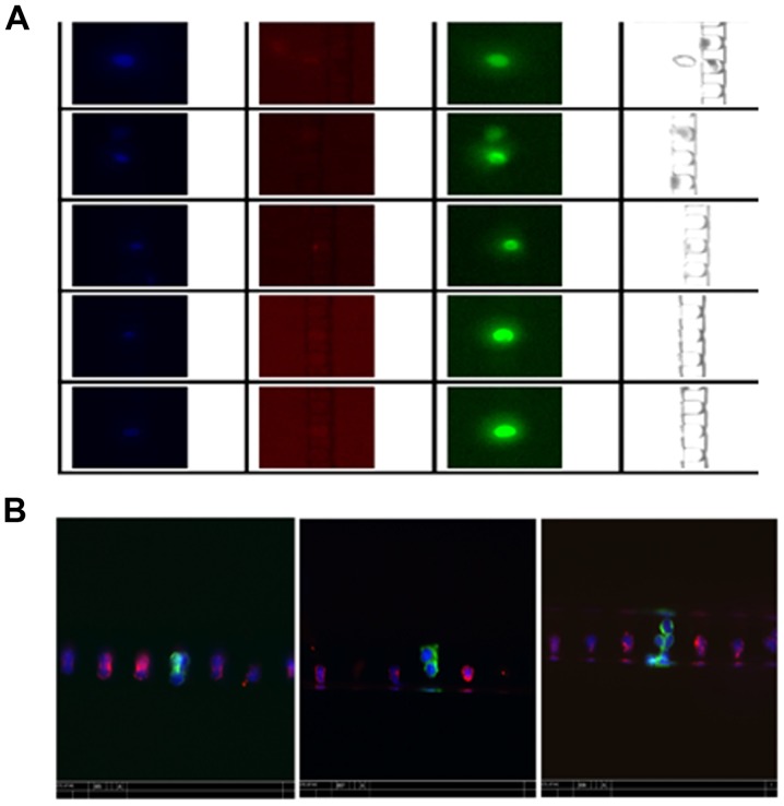
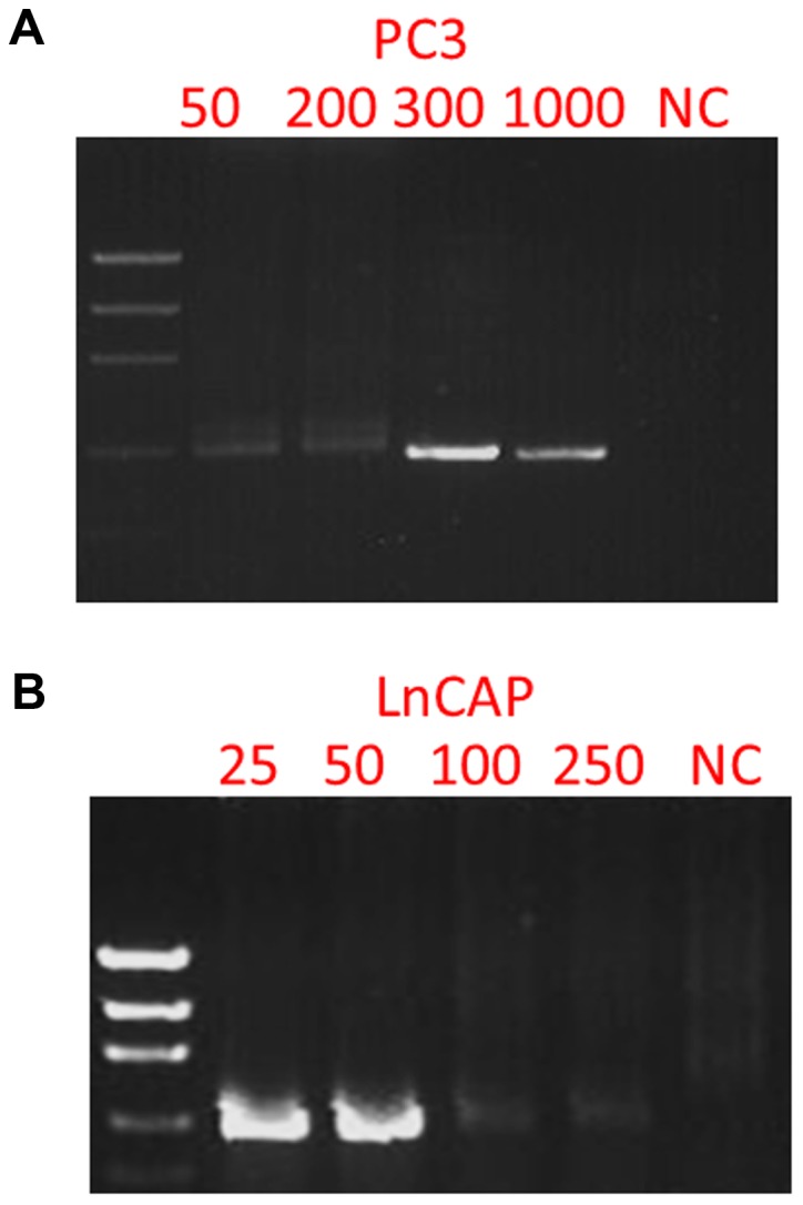
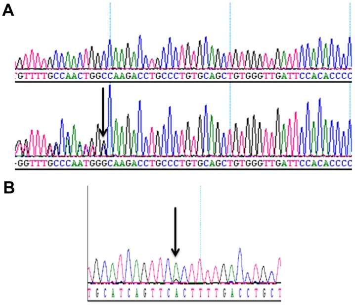
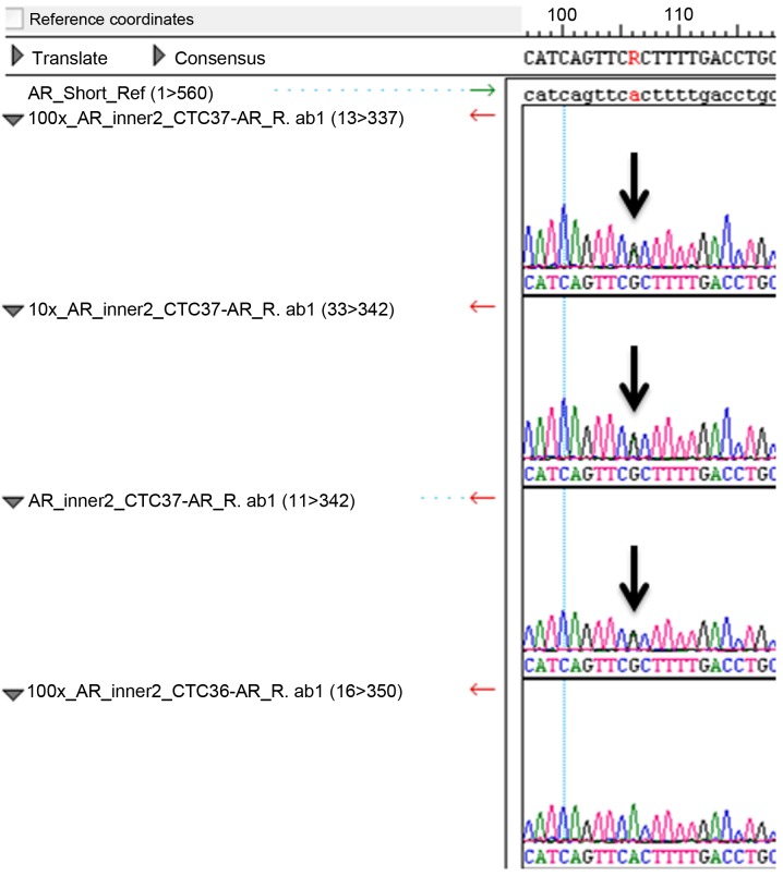
The potential of circulating tumor cells (CTCs) in the diagnosis and prognosis of cancer patients has become increasingly attractive. However, molecular analysis of CTCs is hindered by low sensitivity and a high level of background leukocytes in CTC enrichment technologies. We have developed a novel protocol using a microfluidic device, which enriches and retrieves CTCs from blood samples. The principle of CTC capturing is that tumor cells are larger and less deformable than normal blood cells. To evaluate the potential of utilizing Celsee PREP100 in CTC molecular analysis, we prepared prostate cancer cell lines PC3 and LNCaP, retrieved the captured cells and analyzed them using PCR amplicon sequencing. We were able to recover an average of 79% of 110‑1,100 PC3 and 60‑1,500 LNCaP cells, and detect the p.K139fs*3 deletion of the p53 gene in PC3 cells and p.T877A mutation of the androgen receptor gene in LNCaP cells. Next, we spiked these two types of cells into normal donor blood samples, captured the cells and analyzed them using PCR amplicon sequencing. The PC3 and LNCaP cells were captured and retrieved with the ratio of captured CTCs to the background leukocytes reaching 1:1.5 for PC3 and 1:2.9 for LNCaP cells. We further revealed that the p.K139fs*3 deletion and p.T877A mutation can be detected in the captured PC3 and LNCaP cells, respectively. We successfully validated this approach using clinical blood samples from patients with metastatic prostate cancer. Our results demonstrated a novel approach for CTC enrichment and illustrated the potential of CTC molecular characterization for diagnosis, prognosis and treatment selection of patients with metastatic malignancy.


Figures

Figure 1.
Figure 1.
Nested PCR amplification of the TP53 gene from PC3 cells and the androgen receptor gene from LNCaP cells. (A) Electrophoresis gel imaging of nested PCR amplicons from 0, 28, 280, 400, 800 and 1,200 PC3 cells. (B) Electrophoresis gel imaging of nested PCR amplicons from 0, 5, 10, 20, 25, 100 and 200 LNCaP cells.
Figure 2.
Figure 2.
Sanger sequencing results of the nested PCR amplicons from PC3 and LNCaP cells. (A) Sequencing results of TP53 from control gDNA (upper panel) and PC3 cells (lower panel), revealing a deletion in TP53 from PC3 cells. (B) Sequencing results of AR from control gDNA (upper panel) and LNCaP cells (lower panel), revealing a missense mutation in AR from LNCaP cells.
Figure 3.
Figure 3.
Representative images of the enriched cells using Celsee PREP100. Green, red and blue signals indicate cytokeratins and DAPI-positive, CD45-negative and nuclei, respectively. (A) Images from individual channels. (B) Composite images from all 3 channels revealing differential staining of CTCs (green) and leukocytes (red). It also revealed that circulating tumor cells (CTCs) are captured as a single cell, double cells and cell clusters.
Figure 4.
Figure 4.
PCR products of the TP53 gene from the enriched PC3 cells from blood and the androgen receptor gene from enriched LNCaP cells from blood. (A) Electrophoresis gel imaging of nested PCR amplicons of the enriched cells from blood samples with spiked-in 50, 200, 300, 1,000 PC3 cells. (B) Electrophoresis of nested PCR amplicons of the enriched cells from blood samples with spiked-in 25, 50, 100 and 250 LNCaP cells.
Figure 5.
Figure 5.
Sanger sequencing results of PCR amplicons of the TP53 and androgen receptor genes of the enriched PC3 and LNCaP cells from blood. (A) The upper panel is the sequencing result from the control gDNA and the lower panel is the representative sequencing result of nested PCR amplicons of the enriched PC3 cells. (B) The representative Sanger sequencing result of nested PCR amplicons of the enriched LNCaP cells. Arrows indicate the mutations.
Figure 6.
Figure 6.
Sanger sequencing results of PCR amplicons of the androgen receptor gene of the enriched patient blood samples. The panel is the representative sequencing result of nested PCR amplicons of the patient blood samples (CTC 31 to CTC 41). Sample (CTC 37) carries a heterozygous AR mutation in its circulating tumor cells (CTCs).

References

    1. Pantel K, Alix-Panabières C. Circulating tumor cells in cancer patients: Challenges and perspectives. Trends Mol Med. 2010;16:398–406. doi: 10.1016/j.molmed.2010.07.001. - DOI - PubMed
    1. Maheswaran S, Haber DA. Circulating tumor cells: A window into cancer biology and metastasis. Curr Opin Genet Dev. 2010;20:96–99. doi: 10.1016/j.gde.2009.12.002. - DOI - PMC - PubMed
    1. Cristofanilli M, Budd GT, Ellis MJ, Stopeck A, Matera J, Miller MC, Reuben JM, Doyle GV, Allard WJ, Terstappen LW, et al. Circulating tumor cells, disease progression, and survival in metastatic breast cancer. N Engl J Med. 2004;351:781–791. doi: 10.1056/NEJMoa040766. - DOI - PubMed
    1. Punnoose EA, Atwal SK, Spoerke JM, Savage H, Pandita A, Yeh RF, Pirzkall A, Fine BM, Amler LC, Chen DS, Lackner MR. Molecular biomarker analyses using circulating tumor cells. PLoS One. 2010;5:e12571. doi: 10.1371/journal.pone.0012517. - DOI - PMC - PubMed
    1. Allard WJ, Matera J, Miller MC, Repollet M, Connelly MC, Rao C, Tibbe AG, Uhr JW, Terstappen LW. Tumor cells circulate in the peripheral blood of all major carcinomas but not in healthy subjects or patients with nonmalignant diseases. Clin Cancer Res. 2004;10:6897–6904. doi: 10.1158/1078-0432.CCR-04-0378. - DOI - PubMed