Adult renal neuroblastoma: A case report and literature review
- PMID: 29620664
- PMCID: PMC5902279
- DOI: 10.1097/MD.0000000000010345
Adult renal neuroblastoma: A case report and literature review
Abstract
Rationale: Adult renal neuroblastoma (NB) is extremely rare, and there have been only a few cases previously described in the literature. We report a case of adult renal NB and summarize the clinical and imaging features of the reported cases.
Patient concerns: A 41-year-old female was admitted to our hospital with a chief complaint of gross hematuria that had persisted for a month. Nonenhanced computed tomography (CT) revealed a hypodense right renal mass without calcification. Enhanced CT showed an infiltrative, heterogeneously enhancing right renal mass with retrocaval lymphadenopathy and right renal vein thrombus. Magnetic resonance imaging (MRI) revealed that the right renal mass was isointense relative to the renal parenchyma on nonenhanced T1-weighted images; it showed mixed hypointensity and hyperintensity on T2-weighted images, and heterogeneous enhancement with a hyperintense rim on fat-saturated, enhanced T1W images. The initial impression was renal cell carcinoma (RCC).
Diagnoses: Adult renal neuroblastoma.
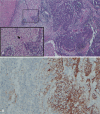
Interventions: Right nephroureterectomy with lymph node dissection was performed. The pathology and immunohistochemistry confirmed the diagnosis of renal NB with retrocaval lymphadenopathy and retroperitoneal metastasis.
Outcomes: After surgery, the patient received 6 courses of chemotherapy, and no recurrence was observed during a 24-month follow-up period.
Lessons: The clinical picture of adult renal NB is that of a 44-year-old woman, presenting with an abdominal or renal mass about 13cm in size, accompanied by hypertension, hematuria, or pain. In contrast to CT features described in previous literature, no tumor calcification is mentioned in these adult renal NB cases. It is difficult to differentiate renal NB from RCC based on CT or MRI. However, biopsy, urinary catecholamine levels, and metaiodobenzylguanidine (MIBG) scan may aid in presurgical diagnosis.
Conflict of interest statement
The authors have no conflicts of interest to disclose.
Figures


References
-
- Franks LM, Bollen A, Seeger RC, et al. Neuroblastoma in adults and adolescents: an indolent course with poor survival. Cancer 1997;79:2028–35. - PubMed
-
- Baumgartner GC, Gaeta J, Wajsman Z, et al. Neuroblastoma presenting as renal cell carcinoma in an adult. Urology 1975;6:376–8. - PubMed
-
- Gohji K, Nakanishi T, Hara I, et al. Two cases of primary neuroblastoma of the kidney in adults. J Urol 1987;137:966–8. - PubMed
-
- Tiu A, Latif E, Yau L, et al. Primary renal neuroblastoma in adults. Urology 2013;82:11–3. - PubMed
Publication types
MeSH terms
LinkOut - more resources
Full Text Sources
Other Literature Sources
Medical

