Review
. 2018 Apr 5;10(4):108.
doi: 10.3390/cancers10040108.

Immunohistochemistry for Diagnosis of Metastatic Carcinomas of Unknown Primary Site

Affiliations
Review

Immunohistochemistry for Diagnosis of Metastatic Carcinomas of Unknown Primary Site

Janick Selves et al. Cancers (Basel). .

Abstract

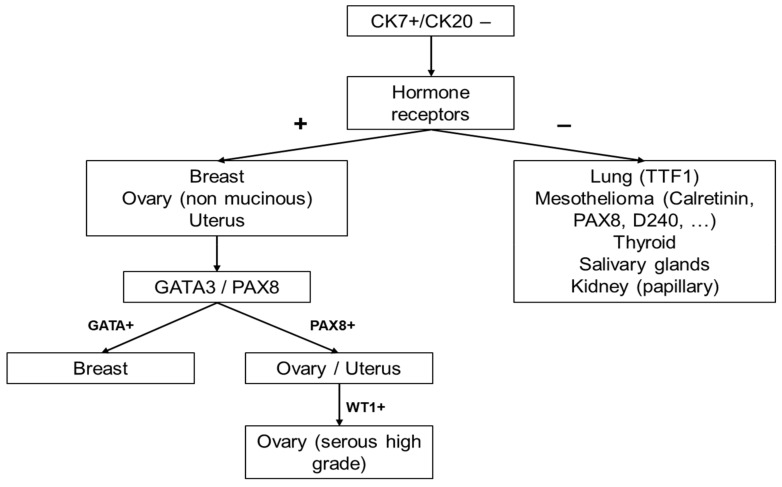
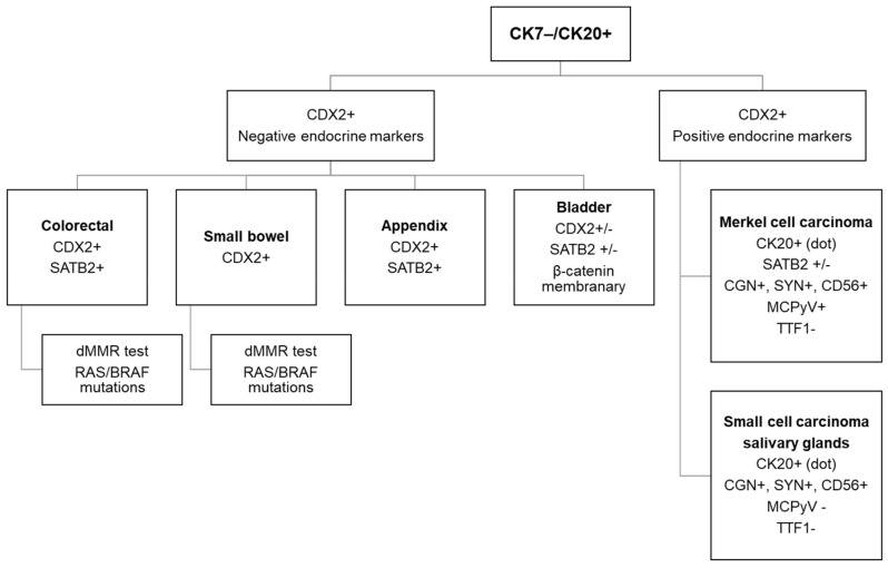
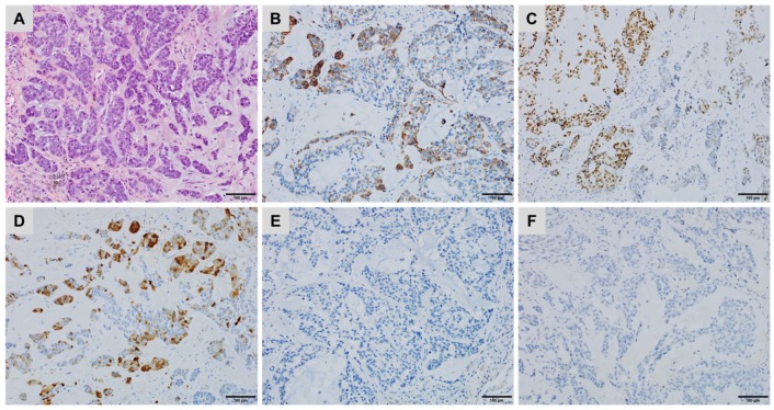
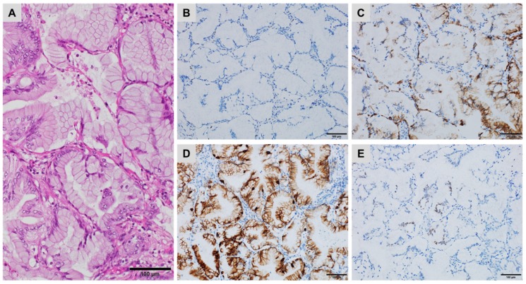
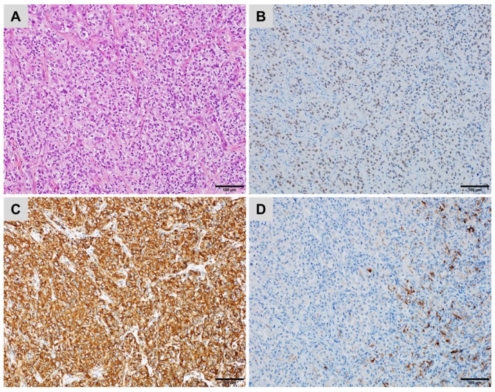
Immunohistochemistry has become an essential ancillary examination for the identification and classification of carcinomas of unknown primary site (CUPs). Over the last decade, the diagnostic accuracy of organ- or tumour-specific immunomarkers and the clinical validation of effective immunohistochemical panels has improved significantly. When dealing with small sample sizes, diagnostic accuracy is crucial, particularly in the current era of targeted molecular and immune-based therapies. Effective systematic use of appropriate immunohistochemical panels enables accurate classification of most of the undifferentiated carcinomas as well as careful preservation of tissues for potential molecular or other ancillary tests. This review discusses the algorithmic approach to the diagnosis of CUPs using CK7 and CK20 staining patterns. It outlines the most frequently used tissue-specific antibodies, provides some pitfalls essential in avoiding potential diagnostic errors and discusses the complementary tools, such as molecular tumour profiling and mutation-specific antibodies, for the improvement of diagnosis and prediction of the treatment response.

Keywords: carcinoma; diagnosis; immunohistochemistry; unknown primary site.


Conflict of interest statement

The authors declare no conflict of interest.

Figures

Figure 1
Figure 1
A lung metastasis of a thyroid carcinoma with a micropapillary component showing (A) cytoplasmic staining for Napsin A, (B) negative staining for Thyroglobulin, (C) diffuse, strong nuclear staining for PAX8 and (D) strong cytoplasmic staining for BRAFV600E (immunoperoxidase, clone VE1).
Figure 2
Figure 2
Pleural effusion showing reactive mesothelial cells mixed with metastatic urothelial carcinoma cells ((A) Giemsa; (B) Papanicolaou staining) demonstrating strong nuclear staining for GATA3 (C).
Figure 3
Figure 3
The diagnostic algorithm in female patients with CK7+/CK20− CUPs.
Figure 4
Figure 4
The diagnostic algorithm for workup of a CK7−/CK20+ carcinoma.
Figure 5
Figure 5
A metastatic urachal adenocarcinoma in the left lung ((A) haematoxylin, eosin and safran) showing patchy staining for (B) CK34βE12, (C) CDX2 and (D) Calretinin but no staining for (E) p40 and (F) GATA3 ((BF) immunoperoxidase).
Figure 6
Figure 6
Primary mucinous pulmonary adenocarcinoma ((A) haematoxylin, eosin and safran) showing (B) no staining for TTF1, (C) patchy staining for CK7, (D) strong staining for CK20 and (E) a few tumour cells stained for CDX2 ((BE) immunoperoxidase).
Figure 7
Figure 7
A metastatic prostatic adenocarcinoma (A) haematoxylin, eosin and safran) showing (B) no staining for prostate-specific antigen (PSA) but (C) diffuse, strong cytoplasmic staining for prostatic specific acid phosphatase (PSAP) ((B,C) immunoperoxidase).
Figure 8
Figure 8
A metastatic renal cell carcinoma ((A) haematoxylin, eosin and safran) showing (B) diffuse nuclear staining for PAX8, (C) diffuse, strong cytoplasmic staining for Vimentin but (D) only a few tumour cells stained for CD10 ((BD) immunoperoxidase).

References

    1. Benderra M.A., Ilie M., Hofman P., Massard C. Standard of care of carcinomas on cancer of unknown primary site in 2016. Bull. Cancer. 2016;103:697–705. doi: 10.1016/j.bulcan.2016.05.003. - DOI - PubMed
    1. Ross J.S., Wang K., Gay L., Otto G.A., White E., Iwanik K., Palmer G., Yelensky R., Lipson D.M., Chmielecki J., et al. Comprehensive genomic profiling of carcinoma of unknown primary site: New routes to targeted therapies. JAMA Oncol. 2015;1:40–49. doi: 10.1001/jamaoncol.2014.216. - DOI - PubMed
    1. Pavlidis N., Pentheroudakis G. Cancer of unknown primary site. Lancet. 2012;379:1428–1435. doi: 10.1016/S0140-6736(11)61178-1. - DOI - PubMed
    1. Bahrami A., Truong L.D., Ro J.Y. Undifferentiated tumor: True identity by immunohistochemistry. Arch. Pathol. Lab. Med. 2008;132:326–348. - PubMed
    1. Greco F.A. Cancer of unknown primary site: Evolving understanding and management of patients. Clin. Adv. Hematol. Oncol. 2012;10:518–524. doi: 10.1016/S1548-5315(11)70301-1. - DOI - PubMed