. 2018 Apr 5;16(4):e2005473.
doi: 10.1371/journal.pbio.2005473. eCollection 2018 Apr.

Ultrastructural localisation of protein interactions using conditionally stable nanobodies

Affiliations

Ultrastructural localisation of protein interactions using conditionally stable nanobodies

Nicholas Ariotti et al. PLoS Biol. .

Abstract

We describe the development and application of a suite of modular tools for high-resolution detection of proteins and intracellular protein complexes by electron microscopy (EM). Conditionally stable GFP- and mCherry-binding nanobodies (termed csGBP and csChBP, respectively) are characterized using a cell-free expression and analysis system and subsequently fused to an ascorbate peroxidase (APEX) enzyme. Expression of these cassettes alongside fluorescently labelled proteins results in recruitment and stabilisation of APEX, whereas unbound APEX nanobodies are efficiently degraded by the proteasome. This greatly simplifies correlative analyses, enables detection of less-abundant proteins, and eliminates the need to balance expression levels between fluorescently labelled and APEX nanobody proteins. Furthermore, we demonstrate the application of this system to bimolecular complementation ('EM split-fluorescent protein'), for localisation of protein-protein interactions at the ultrastructural level.


Conflict of interest statement

The authors have declared that no competing interests exist.

Figures
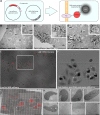
Fig 1
Fig 1. Modular detection of mCherry-tagged proteins using APEX-tagged ChBPs.
A) Schematic of cell-based transfection of modular APEX-ChBP and any mCherry-tagged POI. B-D) Electron micrographs of BHK cells co-expressing APEX-ChBP and B) mCherry, C) mCherry-Cavin1, and D) 2xFYVE-mCherry; arrows highlight areas of enriched electron density. Note the increased density in the cytoplasm compared to mitochondria. Scale bars: lower magnification = 1 μm; insets = 500 nm. E-H) CLEM-based detection nls-mCherry–transfected cells using APEX-ChBP. E) 10x magnification of stacked bright field and epifluorescent images of live BHK cells transfected with H2B-mCherry and APEX-ChBP. The grid coordinate (7K) can be resolved in the bright field image. White box = region of interest. F) Bright field image of flat-embedded cells after removal of the coverslip and tissue culture dish (corresponds to the region of interest from [E]). Significant DAB reaction product can be resolved in the nucleus of cells transfected with the higher expression of the H2B-mCherry. Eight different cells were selected for higher-resolution EM analysis. G) Montaged electron micrographs of the region of interest correlated with red channel epifluorescence image from (E). H) High-resolution transmission electron micrographs of transfected cells (regions 1 to 8, respectively) demonstrated restricted electron density within the nuclei of high-expressing cells (regions 1 to 5) and low-expressing cells (region 6) and no increased electron density above background in untransfected cells (regions 7 and 8). Scale bars: E = 100 μm, F–G = 50 μm, H = 5 μm. DAB, 3,3′-Diaminobenzidine; APEX, ascorbate peroxidase; BHK, baby hamster kidney; Cav, caveolae; CLEM, correlative light and electron microscopy; ChBP, mCherry-binding peptide; Cyto, cytoplasm; EM, electron microscopy; End, endosome; ER, endoplasmic reticulum; H2B, Histone 2B; Mito, Mitochondria; nls-mCherry, nuclear localized mCherry; Nuc, nucleus; PM, plasma membrane; POI, protein of interest.
Fig 2
Fig 2. Conditional stabilisation of GBP and ChBP, and detection of protein–protein interactions using bimolecular fluorescence complementation.
A) Schematic illustrating detection of GFP-tagged POIs using csAPEX-GBP. The probe is degraded by the proteasome unless stabilized by interactions with a GFP-tagged protein, resulting in loss of any nonspecific, electron-dense APEX signal when csAPEX-GBP does not bind to its target. B) csAPEX-GBP shows minimal signal when expressed in cells lacking GFP-tagged proteins; only a low level of labelling is detectable in specific regions of a subset of cells (inset, arrows). In contrast, cells co-expressing soluble GFP together with csAPEX-GBP show a strong cytosolic signal (C, quantitated in S2 Fig. A). D-F) Examples of subcompartment-specific labelling in cells expressing GFP-tagged POIs associating with the PM, the early endosomes, and caveolae, respectively. G-H) Examples of subcompartment-specific labelling in cells expressing mCherry-tagged POIs associating with the PM, nucleus, early endosomes, and caveolae, respectively. K-P) Co-transfection of BHK cells with constructs tagged with each half of split YFP along with csAPEX-GBP gives strong and specific labelling at sites of protein–protein interactions. K) Schematic illustrating detection of interactions between two POIs tagged with different halves of a split YFP. csAPEX-GBP is able to bind only when the YFP pair is fully reconstituted and folded. In the absence of a correctly folded GFP derivative, csAPEX-GBP is degraded by the proteasome. L) Cavin1-YFP-N and Cavin3-YFP-C co-expression gives specific labelling associated with PM pits and vesicular profiles characteristic of caveolae. Note the specificity of the labelling, which allows identification of Cavin1/Cavin3 complexes associated with both surface caveolae and putative endocytic caveolar carriers associated with intracellular compartments (arrow). Further examples are shown in S2B and S2C Fig. M) Reciprocal experimental conditions with specific fragments of YFP switched between constructs gives consistent labelling. N) Cells with an abnormally high transfection level show intracellular aggregates of Cavin (compare with caveolar labelling in L and M). O) Control cells transfected with just one split GFP half and csAPEX-GBP show no labelling in the majority of cells. P) APEX positive inclusions are seen in a small percentage of control cells. These are clearly distinguishable from the specific staining of the recombined protein complex (L-M). Further examples are shown in S2 Fig. D. Scale bars: lower magnification = 1 μm; insets = 500 nm. BHK, baby hamster kidney; CCP, clathrin-coated pits; ChBP, mCherry-binding peptide; cs, conditionally stable; GBP, GFP-nanobody/binding peptide; PM, plasma membrane; POI, protein of interest.

References

    1. Buchfellner A, Yurlova L, Nüske S, Scholz AM, Bogner J, Ruf B, et al. A New Nanobody-Based Biosensor to Study Endogenous PARP1 In Vitro and in Live Human Cells. PLoS ONE. 2016;11(3):e0151041 doi: 10.1371/journal.pone.0151041 - DOI - PMC - PubMed
    1. Tang JC, Drokhlyansky E, Etemad B, Rudolph S, Guo B, Wang S, et al. Detection and manipulation of live antigen-expressing cells using conditionally stable nanobodies. Elife. 2016;5 doi: 10.7554/eLife.15312 - DOI - PMC - PubMed
    1. Lam SS, Martell JD, Kamer KJ, Deerinck TJ, Ellisman MH, Mootha VK, et al. Directed evolution of APEX2 for electron microscopy and proximity labeling. Nature Methods. 2014;1:51–4. doi: 10.1038/nmeth.3179 - DOI - PMC - PubMed
    1. Shi Y, Wang L, Zhang J, Zhai Y, Sun F. Determining the target protein localization in 3D using the combination of FIB-SEM and APEX2. Biophys Rep. 2017;3(4–6):92–9. doi: 10.1007/s41048-017-0043-x - DOI - PMC - PubMed
    1. Ariotti N, Hall TE, Parton RG. Correlative light and electron microscopic detection of GFP-labeled proteins using modular APEX. Methods Cell Biol. 2017;140:105–21. doi: 10.1016/bs.mcb.2017.03.002 - DOI - PubMed

Publication types

MeSH terms

Substances