Review
. 2018 Apr 4;98(1):31-48.
doi: 10.1016/j.neuron.2018.02.022.

Regulation of Body Temperature by the Nervous System

Affiliations
Review

Regulation of Body Temperature by the Nervous System

Chan Lek Tan et al. Neuron. .

Abstract

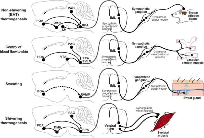
The regulation of body temperature is one of the most critical functions of the nervous system. Here we review our current understanding of thermoregulation in mammals. We outline the molecules and cells that measure body temperature in the periphery, the neural pathways that communicate this information to the brain, and the central circuits that coordinate the homeostatic response. We also discuss some of the key unresolved issues in this field, including the following: the role of temperature sensing in the brain, the molecular identity of the warm sensor, the central representation of the labeled line for cold, and the neural substrates of thermoregulatory behavior. We suggest that approaches for molecularly defined circuit analysis will provide new insight into these topics in the near future.

Keywords: brown-fat thermogenesis; calcium imaging; dorsomedial hypothalamus; neural circuit; optogenetics; preoptic area; shivering; sweating; vasodilation; warm sensor; warm-sensitive neurons.


Figures

Figure 1
Figure 1. Core temperature during challenges to thermal homeostasis
A. Changes in brain or rectal temperatures are typically small during acute external temperature challenges (30-60 minutes) in a range of mammals. B. Changes in brain and rectal temperature in the rat after 30 minutes of exercise (treadmill, average speed 20 m/min, average ambient temp 27°C) or 30 minutes of heat exposure (average 45°C). Solid lines – median C. Rat brain and rectal temperatures are tightly correlated during either exercise or external heating. D. Exercise-induced warming in the rat brain and core are sensitive to prevailing ambient temperatures
Figure 2
Figure 2. Types of thermoregulatory effectors
Examples of physiological and behavioral strategies for controlling body temperature.
Figure 3
Figure 3. The generation of fever
The presence of molecules associated with pathogens like bacteria and viruses is sensed by innate immune cells in the blood and lead to the production of pyrogenic intermediates like cytokines and prostaglandins that act on the preoptic area. In the preoptic area, COX2 expression in endothelial cells result in local PGE2 production, which is the dominant source of fever-inducing PGE2. PGE2 acts through EP3 receptors expressed in the median preoptic (MnPO) to effect changes in body temperature. LPS – lipopolysaccharide; COX2 – cyclooxygenase 2.
Figure 4
Figure 4. Ascending neural pathways that transmit warm and cool signals from the periphery
Structures involved in transmission of thermosensory input from the viscera and skin. Temperature information is sensed by neurons with cell bodies in primary sensory ganglia (or trigeminal ganglia), and then transmitted to the dorsal horn of the spinal cord (or chief sensory nucleus of V), the lateral parabrachial nuclei and finally the preoptic area. Brain regions involved in homeostatic control are shown in gray and those involved in temperature discrimination are shown in blue. Simplified schematics show the responses of neurons in this pathway to external heating and cooling. Sensory ganglia: adapted from (Yarmolinsky et al., 2016). Imaging of neural activity in the trigeminal ganglion shows that over 90% of thermal responsive cells responded to either heating or cooling with 2-5% of cells showing bimodal responses. Upper Line - typical normalized response over 35-50°C tem perature range for the 2 classes of heat sensitive neurons: warmth-sensing neurons with graded responses and broad dynamic range, and noxious heat-sensing neurons with high threshold and narrow dynamic range. Lower plot shows typical responses to cooling to 10°C for the 3 classes of cold-sensing neurons: Type 1 with tonic response to mild-cooling and rapid inactivation by noxious cold; Type 2 with sustained response to noxious cold; and Type 3 with a hybrid response. Spinal cord: adapted from Ran 2016. Imaging of neural activity in dorsal horn showed that cool-active neurons were rapidly adapting and responses scaled with the magnitude of temperature change. Warm-active neurons were non-adapting and responses reflect absolute target temperature. Broadly tuned neurons (not-shown) that responded to both cooling and heating were also present. Lateral parabrachial nucleus: Top – adapted from (Nakamura and Morrison, 2010). Single unit extracellular recording from warm-responsive neurons in the dorsal LPB that project to the preoptic area revealed that activity is increased by skin warming (14 out of 17 cells). Bottom – adapted from (Nakamura and Morrison, 2008). Single unit extracellular recording from cooling-responsive neurons in the external lateral LPB that project to the preoptic area showed that activity is induced in response to skin cooling (11 out of 14). Preoptic area: Adapted from (Tan et al., 2016). Population activity responses of warm-activated PACAP+ neurons in response to external temperature measured by fiber photometry. Preoptic PACAP neurons are progressive activated by increasing temperature from 30 to 42°C. They show no further activity increase in response to noxious heat (>42°C) or activity decrease in response to cold (<30°C).
Figure 5
Figure 5. Interaction between core (brain or spinal cord) and ambient (skin) temperature in the control of thermoregulatory effectors
A and B. Adapted from (Jessen and Ludwig, 1971). Spinal cord (SC) and hypothalamic (Hypo) temperatures in the dog were independently manipulated at varying ambient temperatures and resultant effects of heat production and evaporative heat loss are shown. C. Adapted from (Shafton et al., 2014). Changes in rat tail sympathetic nerve activity (SNA), which is a measure of vasoconstriction (low SNA – vasodilation) as abdominal, skin or brain temperature is altered.
Figure 6
Figure 6. Descending circuits controlling thermoregulatory effectors
The CNS/PNS regions involved invarious thermoregulatory effector responses and the proposed descending pathway from the POA to motor output. Note that many of the connections in the brain that are drawn are postulated based on indirect evidence. Dashed arrows indicate that a functional connection exists, but that the anatomic pathway is unknown and may involve multiple synapses and additional brain regions. POA – preoptic area, DMH – dorsomedial hypothalamus, LH – lateral hypothalamus, PAG – periaqueductal gray, VTA – ventral tegmental area, RMR – raphe medullary region, RPA – raphe pallidus, RVLM – rostral ventrolateral medulla, RVMM – rostral ventromedial medulla, IML – interomediolateral column, SSN – superior salivary nucleus.

References

    1. Abbott SBG, Saper CB. Median preoptic glutamatergic neurons promote thermoregulatory heat loss and water consumption in mice. J Physiol. 2017;595:6569–6583. - PMC - PubMed
    1. Abreu-Vieira G, Xiao C, Gavrilova O, Reitman ML. Integration of body temperature into the analysis of energy expenditure in the mouse. Mol Metab. 2015;4:461–470. - PMC - PubMed
    1. Almeida MC, Hew-Butler T, Soriano RN, Rao S, Wang W, Wang J, Tamayo N, Oliveira DL, Nucci TB, Aryal P, et al. Pharmacological blockade of the cold receptor TRPM8 attenuates autonomic and behavioral cold defenses and decreases deep body temperature. J Neurosci. 2012;32:2086–2099. - PMC - PubMed
    1. Almeida MC, Steiner AA, Branco LG, Romanovsky AA. Neural substrate of cold-seeking behavior in endotoxin shock. PLoS One. 2006;1:e1. - PMC - PubMed
    1. Almeida MC, Vizin RC, Carrettiero DC. Current understanding on the neurophysiology of behavioral thermoregulation. Temperature (Austin) 2015;2:483–490. - PMC - PubMed

Publication types