

Detection of calcium pyrophosphate dihydrate crystals in knee meniscus by dual-energy computed tomography
- PMID: 29622016
- PMCID: PMC5887196
- DOI: 10.1186/s13018-018-0787-0
Detection of calcium pyrophosphate dihydrate crystals in knee meniscus by dual-energy computed tomography
Abstract
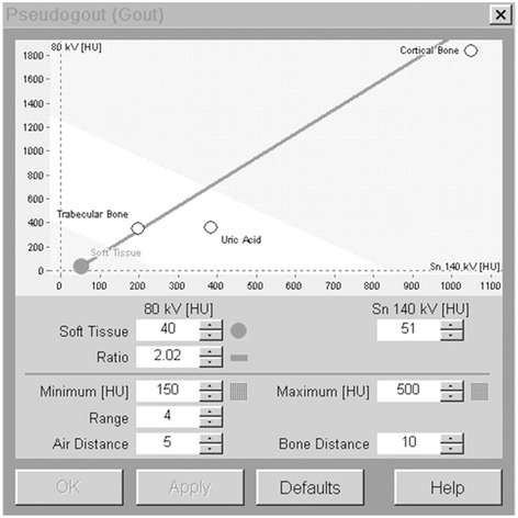
Background: Calcium pyrophosphate dihydrate (CPPD) crystals are commonly observed in osteoarthritic joints. The aim of our study was to investigate the efficacy of a dual-energy computed tomography (DECT) for detecting CPPD crystals in knee meniscus.
Methods: Twenty-six patients undergoing primary total knee arthroplasty were included in the study. Radiographs of knee joint and synovial fluid specimens were analyzed for the presence of CPPD crystals. Meniscus extracted during surgery was scanned using DECT. Sensitivity and specificity of DECT and radiograph for detecting CPPD crystals were calculated against a reference standard (polarizing light microscopy of synovial fluid aspirate). Meniscus in which CPPD crystals were suspected with DECT was further examined to confirm the crystals using a polarized microscopy.
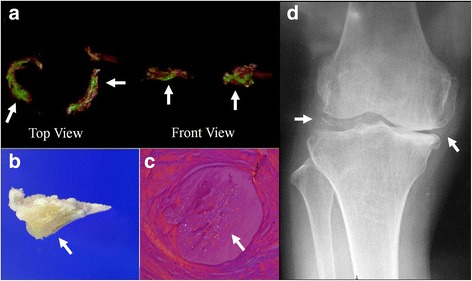
Results: CPPD crystals in synovial fluid were observed in 9 (36%) patients. The sensitivity and specificity of DECT in the detection of CPPD crystals, against microscopic identification, were 77.8 and 93.8%, respectively. The sensitivity and specificity of conventional radiography in the detection of CPPD crystals were 44.4 and 100%, respectively. DECT was able to detect the area where CPPD crystals were deposited in the meniscus.
Conclusion: DECT provides good diagnostic sensitivity and specificity for detection of CPPD crystals in knee meniscus as well as spatial information about CPPD crystals. DECT is currently a research tool, but we believe that DECT can be a useful instrument to diagnose CPPD deposition disease, especially for the regions where aspiration is difficult to be performed such as pubic symphysis, atlantoaxial joint, interphalangeal joint.
Keywords: Calcium pyrophosphate; Dual-energy computed tomography; Knee joint meniscus; Pseudogout.
Conflict of interest statement
Ethics approval and consent to participate
This study followed the Declaration of Helsinki and was approved by the local ethics committee of the Saitama City Hospital, Saitama, Japan.
Consent for publication
Not applicable.
Competing interests
The authors declare that they have no competing interests.
Publisher’s Note
Springer Nature remains neutral with regard to jurisdictional claims in published maps and institutional affiliations.
Figures
References
Publication types
MeSH terms
Substances
LinkOut - more resources
Full Text Sources
Other Literature Sources
